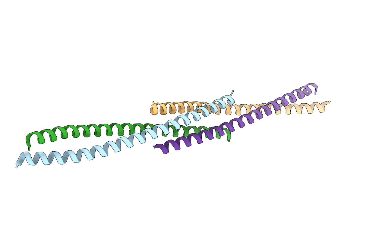
3CVE image
Entry Detail
PDB ID:
3CVE
Title:
Crystal Structure of the carboxy terminus of Homer1
Biological Source:
Source Organism:
Host Organism:
PDB Version:
Deposition Date:
2008-04-18
Release Date:
2009-03-31
Method Details:
Experimental Method:
Resolution:
1.75 Å
R-Value Free:
0.28
R-Value Work:
0.21
R-Value Observed:
0.22
Space Group:
P 1 21 1
Macromolecular Entities
Polymer Type:polypeptide(L)
Description:Homer protein homolog 1
Mutations:L308M
Chain IDs:A, B, C, D
Chain Length:72
Number of Molecules:4
Biological Source:Rattus norvegicus
Modified Residue
Compound ID Chain ID Parent Comp ID Details 2D Image
MSE A MET SELENOMETHIONINE
Primary Citation
The postsynaptic density proteins Homer and Shank form a polymeric network structure.
Cell(Cambridge,Mass.) 137 159 171 (2009)
PMID: 19345194 DOI: 10.1016/j.cell.2009.01.050

Abstact

The postsynaptic density (PSD) is crucial for synaptic functions, but the molecular architecture retaining its structure and components remains elusive. Homer and Shank are among the most abundant scaffolding proteins in the PSD, working synergistically for maturation of dendritic spines. Here, we demonstrate that Homer and Shank, together, form a mesh-like matrix structure. Crystallographic analysis of this region revealed a pair of parallel dimeric coiled coils intercalated in a tail-to-tail fashion to form a tetramer, giving rise to the unique configuration of a pair of N-terminal EVH1 domains at each end of the coiled coil. In neurons, the tetramerization is required for structural integrity of the dendritic spines and recruitment of proteins to synapses. We propose that the Homer-Shank complex serves as a structural framework and as an assembly platform for other PSD proteins.

Legend

Protein

Chemical

Disease

Primary Citation of related structures