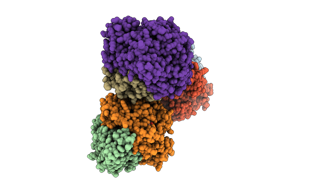
3CUS image
Deposition Date 2008-04-17
Release Date 2008-08-05
Last Version Date 2024-11-20
Entry Detail
PDB ID:
3CUS
Keywords:
Title:
Structure of a double ILE/PHE mutant of NI-FE hydrogenase refined at 2.2 angstrom resolution
Biological Source:
Source Organism:
Method Details:
Experimental Method:
Resolution:
2.20 Å
R-Value Free:
0.22
R-Value Work:
0.18
R-Value Observed:
0.18
Space Group:
P 1 21 1
Macromolecular Entities
Polymer Type:polypeptide(L)
Molecule:Periplasmic [NiFe] hydrogenase small subunit
Gene (Uniprot):hydA
Chain IDs:A, C (auth: B), E (auth: C)
Chain Length:264
Number of Molecules:3
Biological Source:Desulfovibrio fructosovorans
Polymer Type:polypeptide(L)
Molecule:Periplasmic [NiFe] hydrogenase large subunit
Gene (Uniprot):hydB
Mutations:V74I, L122F
Chain IDs:B (auth: Q), D (auth: R), F (auth: S)
Chain Length:549
Number of Molecules:3
Biological Source:Desulfovibrio fructosovorans
Modified Residue
Compound ID Chain ID Parent Comp ID Details 2D Image
CSO B CYS S-HYDROXYCYSTEINE
Primary Citation

Abstact

Hydrogenases, which catalyze H(2) to H(+) conversion as part of the bioenergetic metabolism of many microorganisms, are among the metalloenzymes for which a gas-substrate tunnel has been described by using crystallography and molecular dynamics. However, the correlation between protein structure and gas-diffusion kinetics is unexplored. Here, we introduce two quantitative methods for probing the rates of diffusion within hydrogenases. One uses protein film voltammetry to resolve the kinetics of binding and release of the competitive inhibitor CO; the other is based on interpreting the yield in the isotope exchange assay. We study structurally characterized mutants of a NiFe hydrogenase, and we show that two mutations, which significantly narrow the tunnel near the entrance of the catalytic center, decrease the rates of diffusion of CO and H(2) toward and from the active site by up to 2 orders of magnitude. This proves the existence of a functional channel, which matches the hydrophobic cavity found in the crystal. However, the changes in diffusion rates do not fully correlate with the obstruction induced by the mutation and deduced from the x-ray structures. Our results demonstrate the necessity of measuring diffusion rates and emphasize the role of side-chain dynamics in determining these.

Legend

Protein

Chemical

Disease

Primary Citation of related structures