Deposition Date
2008-04-14
Release Date
2008-07-29
Last Version Date
2024-02-21
Entry Detail
PDB ID:
3CTR
Keywords:
Title:
Crystal structure of the RRM-domain of the poly(A)-specific ribonuclease PARN bound to m7GTP
Biological Source:
Source Organism(s):
Homo sapiens (Taxon ID: 9606)
Expression System(s):
Method Details:
Experimental Method:
Resolution:
2.10 Å
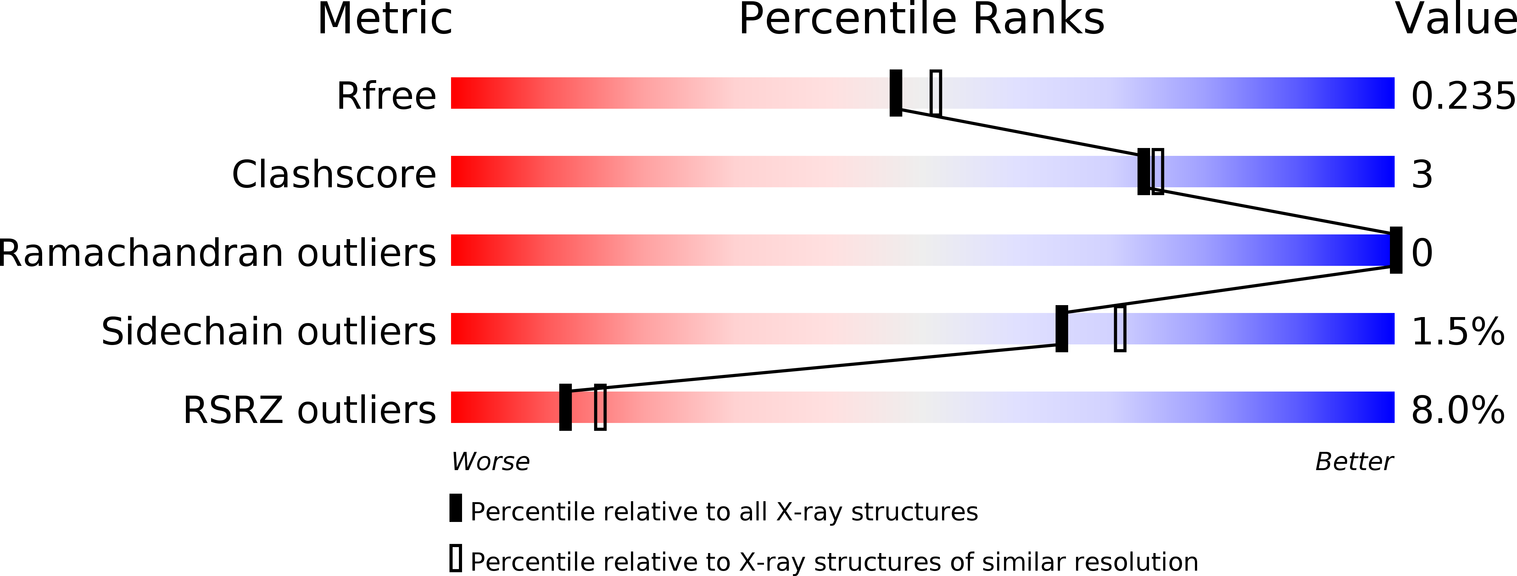
R-Value Free:
0.24
R-Value Work:
0.20
R-Value Observed:
0.21
Space Group:
I 41 2 2