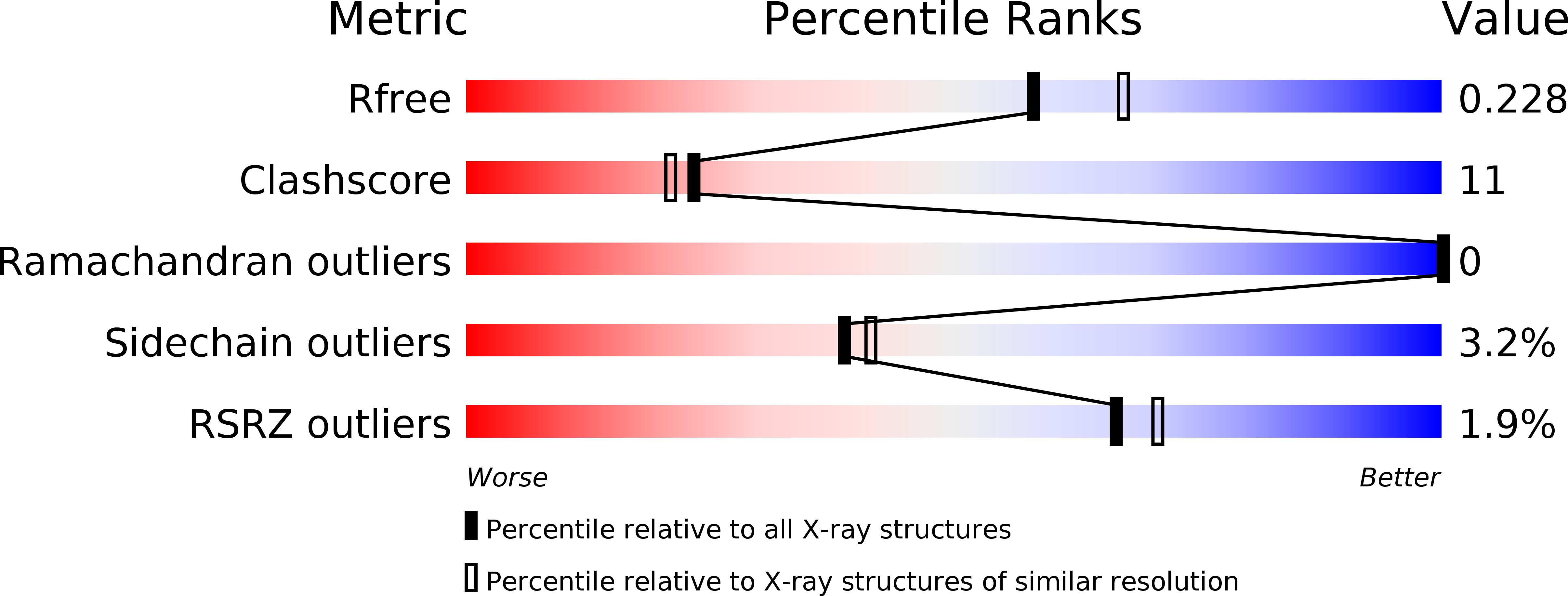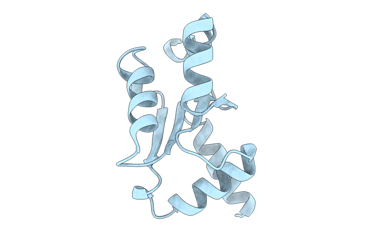
3CTF image
Deposition Date 2008-04-14
Release Date 2008-11-11
Last Version Date 2024-11-06
Entry Detail
PDB ID:
3CTF
Keywords:
Title:
Crystal structure of oxidized GRX2
Biological Source:
Source Organism:
Host Organism:
Method Details:
Experimental Method:
Resolution:
2.10 Å
R-Value Free:
0.25
R-Value Work:
0.22
R-Value Observed:
0.22
Space Group:
P 41 21 2
Macromolecular Entities
Polymer Type:polypeptide(L)
Molecule:Glutaredoxin-2
Gene (Uniprot):GRX2
Chain IDs:A
Chain Length:129
Number of Molecules:1
Biological Source:Saccharomyces cerevisiae
Primary Citation
Structural basis for the different activities of yeast Grx1 and Grx2.
Biochim.Biophys.Acta 1804 1542 1547 (2010)
PMID: 20417731 DOI: 10.1016/j.bbapap.2010.04.010

Abstact

Yeast glutaredoxins Grx1 and Grx2 catalyze the reduction of both inter- and intra-molecular disulfide bonds using glutathione (GSH) as the electron donor. Although sharing the same dithiolic CPYC active site and a sequence identity of 64%, they have been proved to play different roles during oxidative stress and to possess different glutathione-disulfide reductase activities. To address the structural basis of these differences, we solved the crystal structures of Grx2 in oxidized and reduced forms, at 2.10 A and 1.50 A, respectively. With the Grx1 structures we previously reported, comparative structural analyses revealed that Grx1 and Grx2 share a similar GSH binding site, except for a single residue substitution from Asp89 in Grx1 to Ser123 in Grx2. Site-directed mutagenesis in combination with activity assays further proved this single residue variation is critical for the different activities of yeast Grx1 and Grx2.

Legend

Protein

Chemical

Disease

Primary Citation of related structures