Deposition Date
2008-04-10
Release Date
2008-06-10
Last Version Date
2024-02-21
Entry Detail
PDB ID:
3CSX
Keywords:
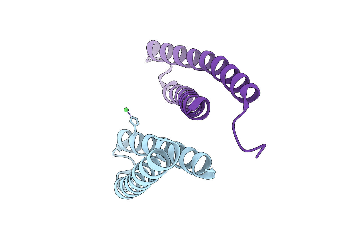
Title:
Structural characterization of a protein in the DUF683 family- crystal structure of cce_0567 from the cyanobacterium Cyanothece 51142.
Biological Source:
Source Organism(s):
Cyanothece (Taxon ID: )
Expression System(s):
Method Details:
Experimental Method:
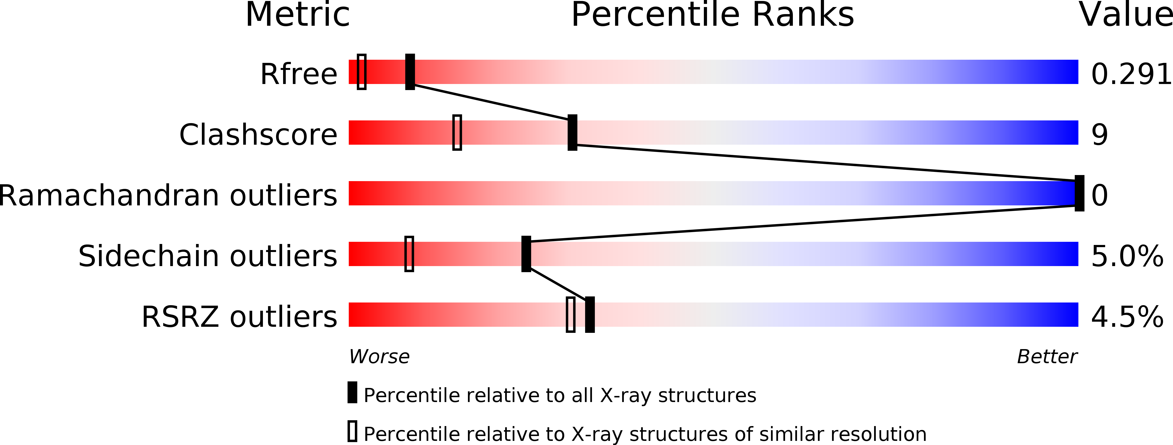
Resolution:
1.84 Å
R-Value Free:
0.29
R-Value Work:
0.21
R-Value Observed:
0.21
Space Group:
P 1 21 1