
Deposition Date
2008-04-10
Release Date
2008-04-22
Last Version Date
2023-08-30
Entry Detail
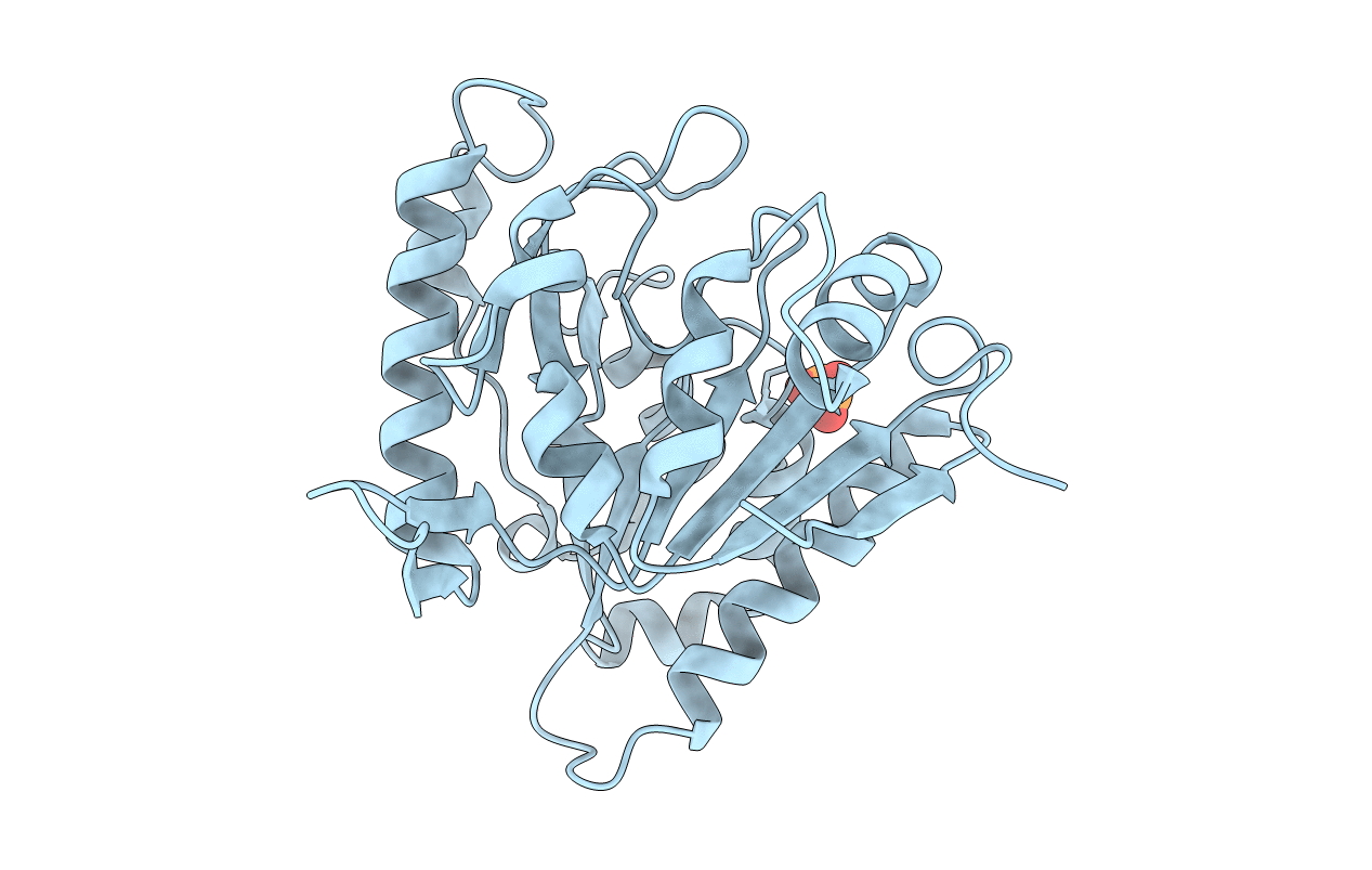
PDB ID:
3CSS
Keywords:
Title:
Crystal structure of 6-phosphogluconolactonase from Leishmania guyanensis
Biological Source:
Source Organism(s):
Leishmania braziliensis (Taxon ID: 420245)
Expression System(s):
Method Details:
Experimental Method:
Resolution:
1.70 Å
R-Value Free:
0.18
R-Value Work:
0.16
R-Value Observed:
0.17
Space Group:
C 2 2 21
