
Deposition Date
2008-04-10
Release Date
2009-01-20
Last Version Date
2024-10-16
Entry Detail
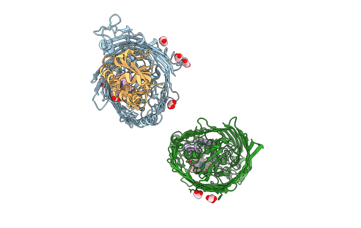
PDB ID:
3CSL
Keywords:
Title:
Structure of the Serratia marcescens hemophore receptor HasR in complex with its hemophore HasA and heme
Biological Source:
Source Organism(s):
Serratia marcescens (Taxon ID: 615)
Expression System(s):
Method Details:
Experimental Method:
Resolution:
2.70 Å
R-Value Free:
0.27
R-Value Work:
0.23
R-Value Observed:
0.24
Space Group:
F 2 2 2
