3CMQ image
Deposition Date 2008-03-24
Release Date 2008-07-01
Last Version Date 2023-08-30
Entry Detail
PDB ID:
3CMQ
Keywords:
Title:
Crystal structure of human mitochondrial phenylalanine tRNA synthetase
Biological Source:
Source Organism:
Homo sapiens (Taxon ID: )
Host Organism:
Method Details:
Experimental Method:
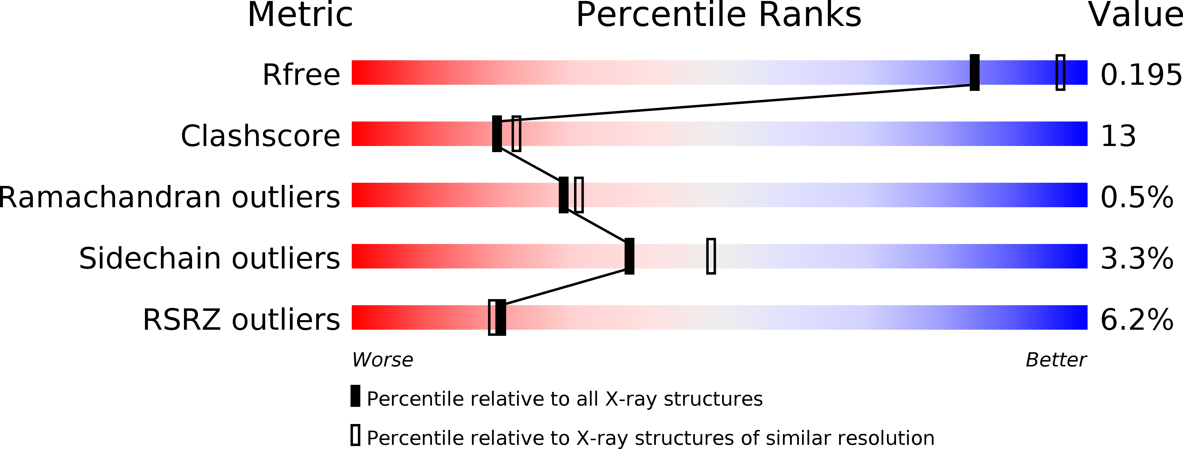
Resolution:
2.20 Å
R-Value Free:
0.24
R-Value Work:
0.19
R-Value Observed:
0.24
Space Group:
P 21 21 21
Macromolecular Entities
Structures with similar UniProt ID
Protein Blast
Polymer Type:polypeptide(L)
Molecule:Phenylalanyl-tRNA synthetase, mitochondrial
Gene (Uniprot):FARS2
Chain IDs:A
Chain Length:415
Number of Molecules:1
Biological Source:Homo sapiens
Primary Citation
The tRNA-Induced Conformational Activation of Human Mitochondrial Phenylalanyl-tRNA Synthetase.
Structure 16 1095 1104 (2008)
PMID: 18611382 DOI: 10.1016/j.str.2008.03.020

Abstact

All class II aminoacyl-tRNA synthetases (aaRSs) are known to be active as functional homodimers, homotetramers, or heterotetramers. However, multimeric organization is not a prerequisite for phenylalanylation activity, as monomeric mitochondrial phenylalanyl-tRNA synthetase (PheRS) is also active. We herein report the structure, at 2.2 A resolution, of a human monomeric mitPheRS complexed with Phe-AMP. The smallest known aaRS, which is, in fact, 1/5 of a cytoplasmic analog, is a chimera of the catalytic module of the alpha and anticodon binding domain (ABD) of the bacterial beta subunit of (alphabeta)2 PheRS. We demonstrate that the ABD located at the C terminus of mitPheRS overlaps with the acceptor stem of phenylalanine transfer RNA (tRNAPhe) if the substrate is positioned in a manner similar to that seen in the binary Thermus thermophilus complex. Thus, formation of the PheRS-tRNAPhe complex in human mitochondria must be accompanied by considerable rearrangement (hinge-type rotation through approximately 160 degrees) of the ABD upon tRNA binding.

Legend

Protein

Chemical

Disease

Primary Citation of related structures