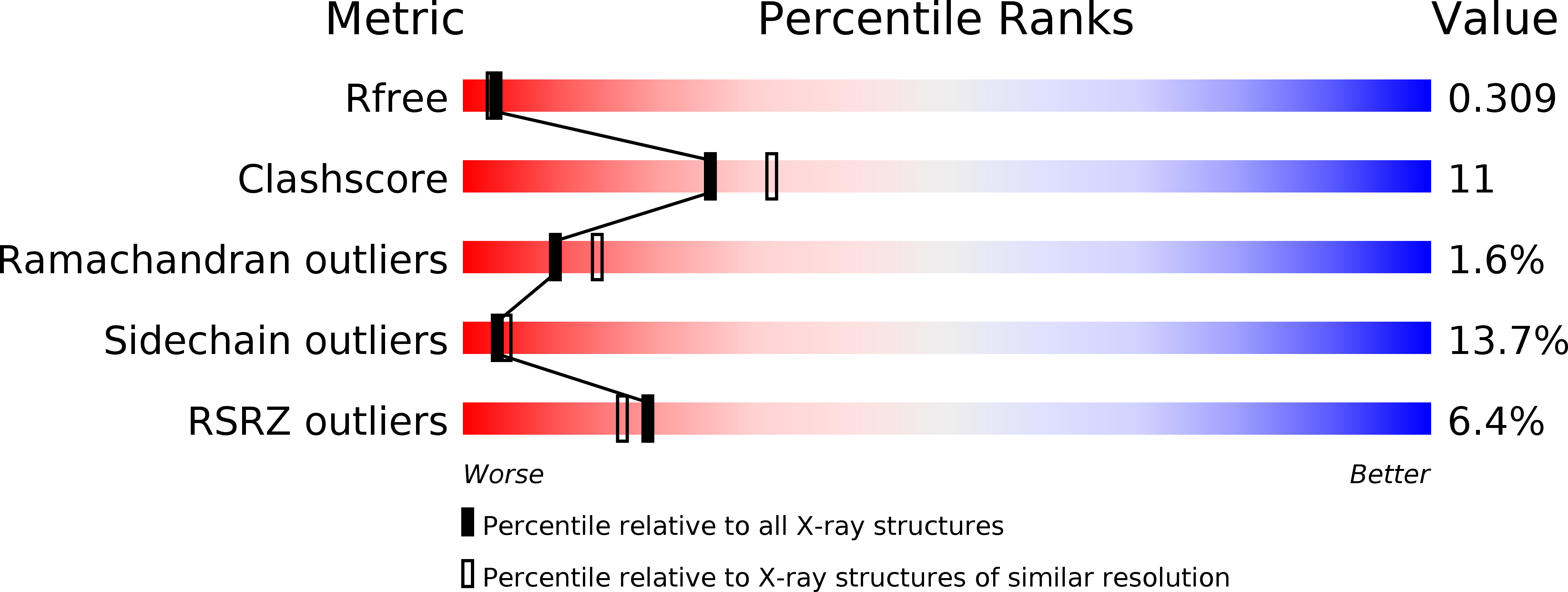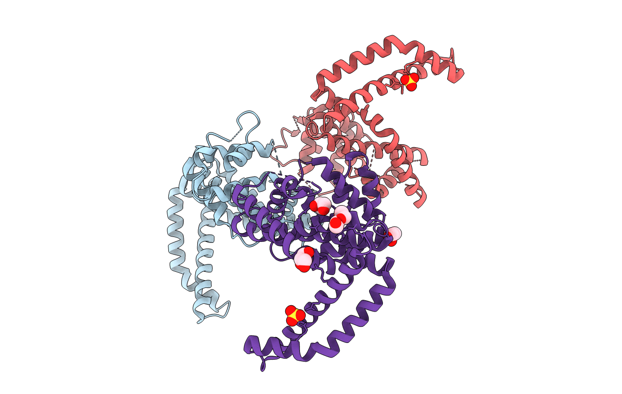
3CKD image
Entry Detail
PDB ID:
3CKD
Keywords:
Title:
Crystal structure of the C-terminal domain of the Shigella type III effector IpaH
Biological Source:
Host Organism:
PDB Version:
Deposition Date:
2008-03-14
Release Date:
2008-03-25
Method Details:
Experimental Method:
Resolution:
2.65 Å
R-Value Free:
0.28
R-Value Work:
0.22
R-Value Observed:
0.23
Space Group:
I 4 2 2
Macromolecular Entities
Polymer Type:polypeptide(L)
Description:Invasion plasmid antigen, secreted by the Mxi-Spa secretion machinery
Chain IDs:A, B, C
Chain Length:312
Number of Molecules:3
Biological Source:Shigella flexneri 2a str. 301
Modified Residue
Compound ID Chain ID Parent Comp ID Details 2D Image
MSE A MET SELENOMETHIONINE
Primary Citation
Structure of the Shigella T3SS effector IpaH defines a new class of E3 ubiquitin ligases.
Nat.Struct.Mol.Biol. 15 1293 1301 (2008)
PMID: 18997778 DOI: 10.1038/nsmb.1511

Abstact

IpaH proteins are E3 ubiquitin ligases delivered by the type III secretion apparatus into host cells upon infection of humans by the Gram-negative pathogen Shigella flexneri. These proteins comprise a variable leucine-rich repeat-containing N-terminal domain and a conserved C-terminal domain harboring an invariant cysteine residue that is crucial for activity. IpaH homologs are encoded by diverse animal and plant pathogens. Here we demonstrate that the IpaH C-terminal domain carries the catalytic activity for ubiquitin transfer and that the N-terminal domain carries the substrate specificity. The structure of the IpaH C-terminal domain, determined to 2.65-A resolution, represents an all-helical fold bearing no resemblance to previously defined E3 ubiquitin ligases. The conserved and essential cysteine residue lies on a flexible, surface-exposed loop surrounded by conserved acidic residues, two of which are crucial for IpaH activity.

Legend

Protein

Chemical

Disease

Primary Citation of related structures