3CK9 image
Deposition Date 2008-03-14
Release Date 2008-05-20
Last Version Date 2023-08-30
Entry Detail
PDB ID:
3CK9
Title:
B. thetaiotaomicron SusD with maltoheptaose
Biological Source:
Source Organism:
Host Organism:
Method Details:
Experimental Method:
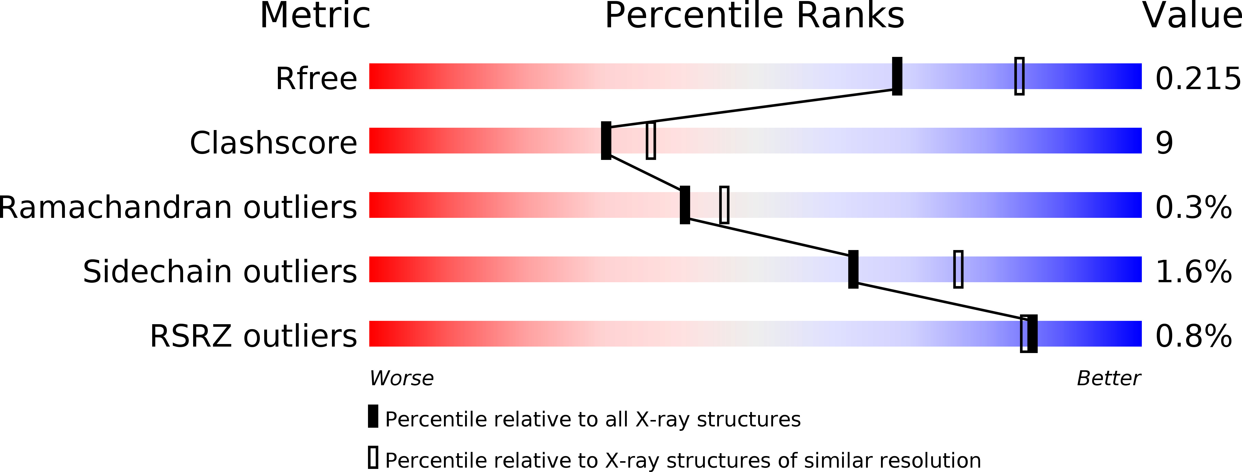
Resolution:
2.20 Å
R-Value Free:
0.22
R-Value Work:
0.18
R-Value Observed:
0.18
Space Group:
P 1
Macromolecular Entities
Polymer Type:polypeptide(L)
Molecule:SusD
Gene (Uniprot):susD
Chain IDs:A, B
Chain Length:527
Number of Molecules:2
Biological Source:Bacteroides thetaiotaomicron
Peptide-like Molecules
PRD_900035
Primary Citation
Starch catabolism by a prominent human gut symbiont is directed by the recognition of amylose helices.
Structure 16 1105 1115 (2008)
PMID: 18611383 DOI: 10.1016/j.str.2008.03.017

Abstact

The human gut microbiota performs functions that are not encoded in our Homo sapiens genome, including the processing of otherwise undigestible dietary polysaccharides. Defining the structures of proteins involved in the import and degradation of specific glycans by saccharolytic bacteria complements genomic analysis of the nutrient-processing capabilities of gut communities. Here, we describe the atomic structure of one such protein, SusD, required for starch binding and utilization by Bacteroides thetaiotaomicron, a prominent adaptive forager of glycans in the distal human gut microbiota. The binding pocket of this unique alpha-helical protein contains an arc of aromatic residues that complements the natural helical structure of starch and imposes this conformation on bound maltoheptaose. Furthermore, SusD binds cyclic oligosaccharides with higher affinity than linear forms. The structures of several SusD/oligosaccharide complexes reveal an inherent ligand recognition plasticity dominated by the three-dimensional conformation of the oligosaccharides rather than specific interactions with the composite sugars.

Legend

Protein

Chemical

Disease

Primary Citation of related structures