Deposition Date
2008-03-13
Release Date
2009-03-24
Last Version Date
2024-11-20
Entry Detail
PDB ID:
3CJJ
Keywords:
Title:
Crystal structure of human rage ligand-binding domain
Biological Source:
Source Organism(s):
Homo sapiens (Taxon ID: 9606)
Expression System(s):
Method Details:
Experimental Method:
Resolution:
1.85 Å
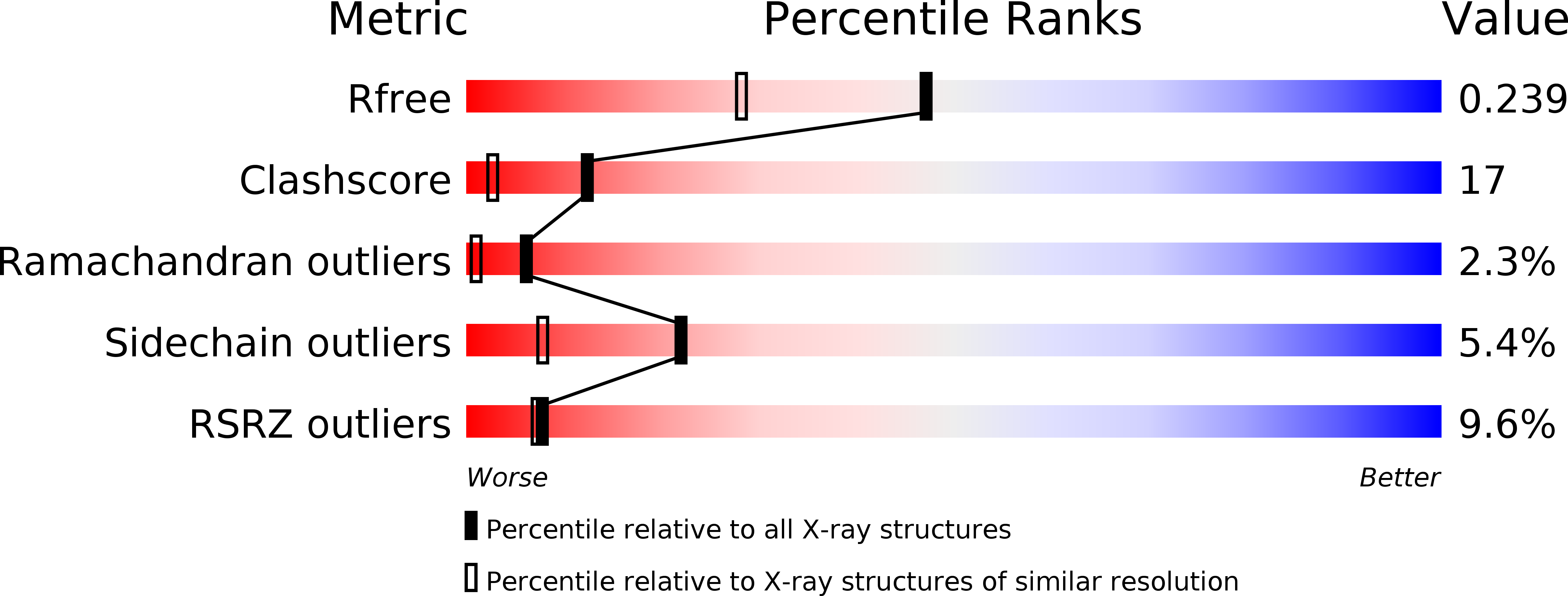
R-Value Free:
0.24
R-Value Work:
0.20
R-Value Observed:
0.21
Space Group:
P 21 21 2