3CIO image
Entry Detail
PDB ID:
3CIO
Title:
The kinase domain of Escherichia coli tyrosine kinase ETK
Biological Source:
Source Organism:
Host Organism:
PDB Version:
Deposition Date:
2008-03-11
Release Date:
2008-06-03
Method Details:
Experimental Method:
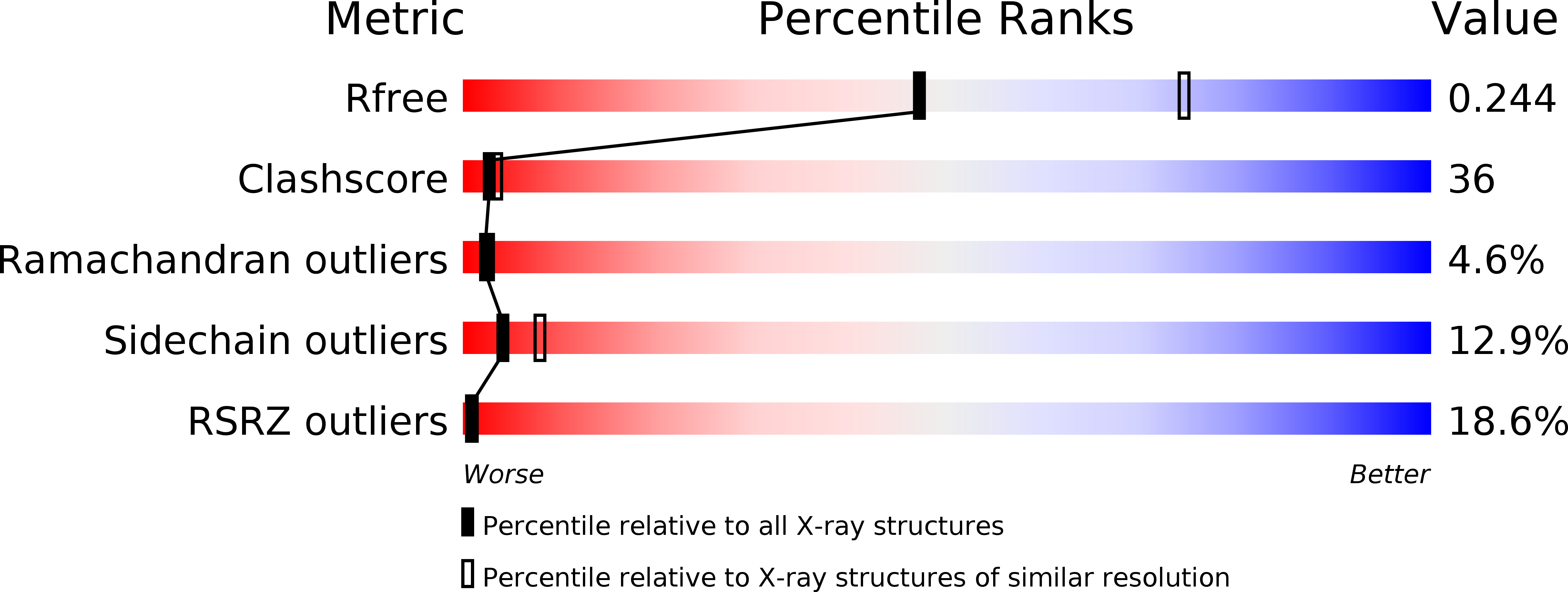
Resolution:
2.50 Å
R-Value Free:
0.25
R-Value Work:
0.19
R-Value Observed:
0.19
Space Group:
C 1 2 1
Macromolecular Entities
Polymer Type:polypeptide(L)
Description:Tyrosine-protein kinase etk
Chain IDs:A, B (auth: D)
Chain Length:299
Number of Molecules:2
Biological Source:Escherichia coli
Ligand Molecules
Primary Citation
Structure of Escherichia coli tyrosine kinase Etk reveals a novel activation mechanism.
Embo J. 27 1758 1766 (2008)
PMID: 18497741 DOI: 10.1038/emboj.2008.97

Abstact

While protein tyrosine (Tyr) kinases (PTKs) have been extensively characterized in eukaryotes, far less is known about their emerging counterparts in prokaryotes. The inner-membrane Wzc/Etk protein belongs to the bacterial PTK family, which has an important function in regulating the polymerization and transport of virulence-determining capsular polysaccharide (CPS). The kinase uses a unique two-step activation process involving intra-phosphorylation of a Tyr residue, although the molecular mechanism remains unknown. Herein, we report the first crystal structure of a bacterial PTK, the C-terminal kinase domain of Escherichia coli Tyr kinase (Etk) at 2.5-A resolution. The fold of the Etk kinase domain differs markedly from that of eukaryotic PTKs. Based on the observed structure and supporting mass spectrometric evidence of Etk, a unique activation mechanism is proposed that involves the phosphorylated Tyr residue, Y574, at the active site and its specific interaction with a previously unidentified key Arg residue, R614, to unblock the active site. Both in vitro kinase activity and in vivo antibiotics resistance studies using structure-guided mutants further support the novel activation mechanism.

Legend

Protein

Chemical

Disease

Primary Citation of related structures