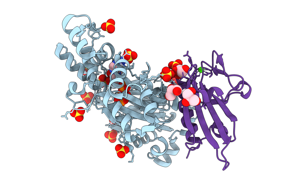
3CI5 image
Deposition Date 2008-03-10
Release Date 2008-08-19
Last Version Date 2023-11-15
Entry Detail
PDB ID:
3CI5
Title:
Complex of Phosphorylated Dictyostelium Discoideum Actin with Gelsolin
Biological Source:
Source Organism:
Homo sapiens (Taxon ID: )
Host Organism:
Method Details:
Experimental Method:
Resolution:
1.70 Å
R-Value Free:
0.19
R-Value Work:
0.14
R-Value Observed:
0.14
Space Group:
C 1 2 1
Macromolecular Entities
Polymer Type:polypeptide(L)
Molecule:Major actin
Gene (Uniprot):act1, act2, act4, act5, act6, act7, act8, act9, act11, act12, act13, act14, act15, act16, act19, act20, act21
Chain IDs:A
Chain Length:375
Number of Molecules:1
Biological Source:Dictyostelium discoideum
Polymer Type:polypeptide(L)
Molecule:Gelsolin
Gene (Uniprot):GSN
Chain IDs:B (auth: G)
Chain Length:126
Number of Molecules:1
Biological Source:Homo sapiens
Modified Residue
Compound ID Chain ID Parent Comp ID Details 2D Image
HIC A HIS 4-METHYL-HISTIDINE
PTR A TYR O-PHOSPHOTYROSINE
Primary Citation
Modulation of actin structure and function by phosphorylation of Tyr-53 and profilin binding.
Proc.Natl.Acad.Sci.Usa 105 11748 11753 (2008)
PMID: 18689676 DOI: 10.1073/pnas.0805852105

Abstact

On starvation, Dictyostelium cells aggregate to form multicellular fruiting bodies containing spores that germinate when transferred to nutrient-rich medium. This developmental cycle correlates with the extent of actin phosphorylation at Tyr-53 (pY53-actin), which is low in vegetative cells but high in viable mature spores. Here we describe high-resolution crystal structures of pY53-actin and unphosphorylated actin in complexes with gelsolin segment 1 and profilin. In the structure of pY53-actin, the phosphate group on Tyr-53 makes hydrogen-bonding interactions with residues of the DNase I-binding loop (D-loop) of actin, resulting in a more stable conformation of the D-loop than in the unphosphorylated structures. A more rigidly folded D-loop may explain some of the previously described properties of pY53-actin, including its increased critical concentration for polymerization, reduced rates of nucleation and pointed end elongation, and weak affinity for DNase I. We show here that phosphorylation of Tyr-53 inhibits subtilisin cleavage of the D-loop and reduces the rate of nucleotide exchange on actin. The structure of profilin-Dictyostelium-actin is strikingly similar to previously determined structures of profilin-beta-actin and profilin-alpha-actin. By comparing this representative set of profilin-actin structures with other structures of actin, we highlight the effects of profilin on the actin conformation. In the profilin-actin complexes, subdomains 1 and 3 of actin close around profilin, producing a 4.7 degrees rotation of the two major domains of actin relative to each other. As a result, the nucleotide cleft becomes moderately more open in the profilin-actin complex, probably explaining the stimulation of nucleotide exchange on actin by profilin.

Legend

Protein

Chemical

Disease

Primary Citation of related structures