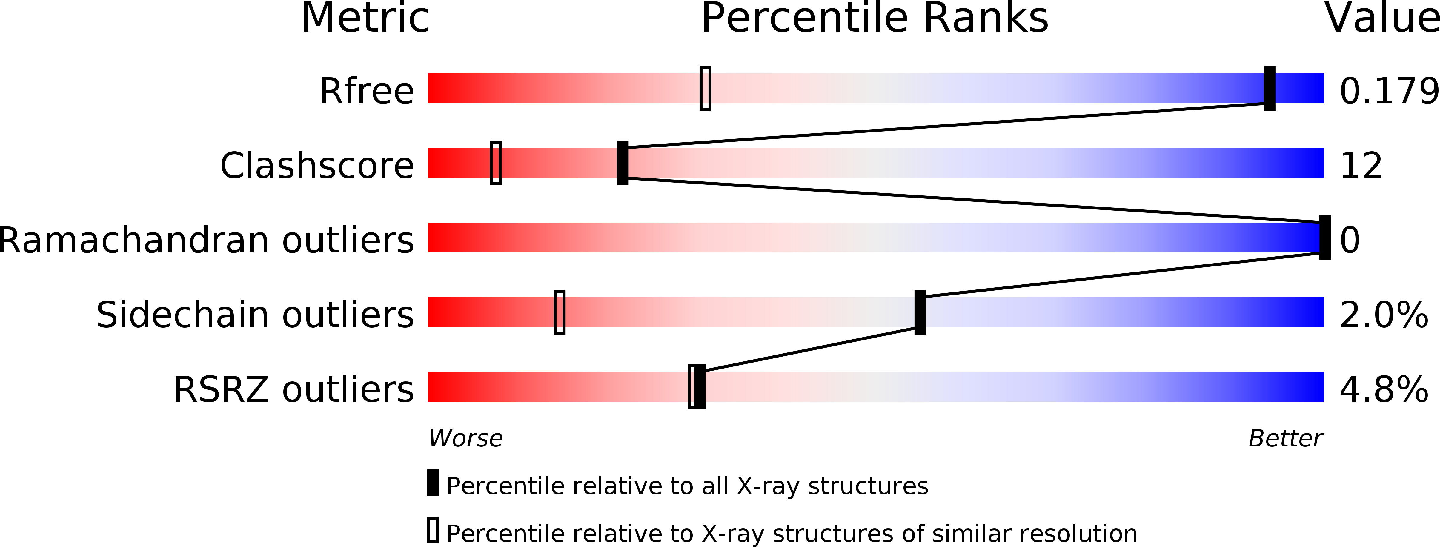
3CI3 image
Deposition Date 2008-03-10
Release Date 2008-05-27
Last Version Date 2024-02-21
Entry Detail
PDB ID:
3CI3
Keywords:
Title:
Structure of the PduO-type ATP:co(I)rrinoid adenosyltransferase from Lactobacillus reuteri complexed with partial adenosylcobalamin and PPPi
Biological Source:
Source Organism:
Host Organism:
Method Details:
Experimental Method:
Resolution:
1.11 Å
R-Value Free:
0.18
R-Value Work:
0.16
R-Value Observed:
0.16
Space Group:
H 3
Macromolecular Entities
Polymer Type:polypeptide(L)
Molecule:Cobalamin adenosyltransferase PduO-like protein
Gene (Uniprot):cobA
Chain IDs:A
Chain Length:194
Number of Molecules:1
Biological Source:Lactobacillus reuteri
Primary Citation
Structural characterization of a human-type corrinoid adenosyltransferase confirms that coenzyme B12 is synthesized through a four-coordinate intermediate.
Biochemistry 47 5755 5766 (2008)
PMID: 18452306 DOI: 10.1021/bi800132d

Abstact

ATP:cob(I)alamin adenosyltransferases (ACAs) catalyze the transfer of the 5'-deoxyadenosyl moiety from ATP to the upper axial ligand position of cobalamin in the synthesis of coenzyme B 12. For the ACA-catalyzed reaction to proceed, cob(II)alamin must be reduced to cob(I)alamin in the enzyme active site. This reduction is facilitated through the generation of a four-coordinate cob(II)alamin intermediate on the enzyme. We have determined the high-resolution crystal structure of a human-type ACA from Lactobacillus reuteri with a four-coordinate cob(II)alamin bound in the enzyme active site and with the product, adenosylcobalamin, partially occupied in the active site. The assembled structures represent snapshots of the steps in the ACA-catalyzed formation of the cobalt-carbon bond of coenzyme B 12. The structures define the corrinoid binding site and provide visual evidence for a base-off, four-coordinate cob(II)alamin intermediate. The complete structural description of ACA-mediated catalysis reveals the molecular features of four-coordinate cob(II)alamin stabilization and provides additional insights into the molecular basis for dysfunction in human patients suffering from methylmalonic aciduria.

Legend

Protein

Chemical

Disease

Primary Citation of related structures