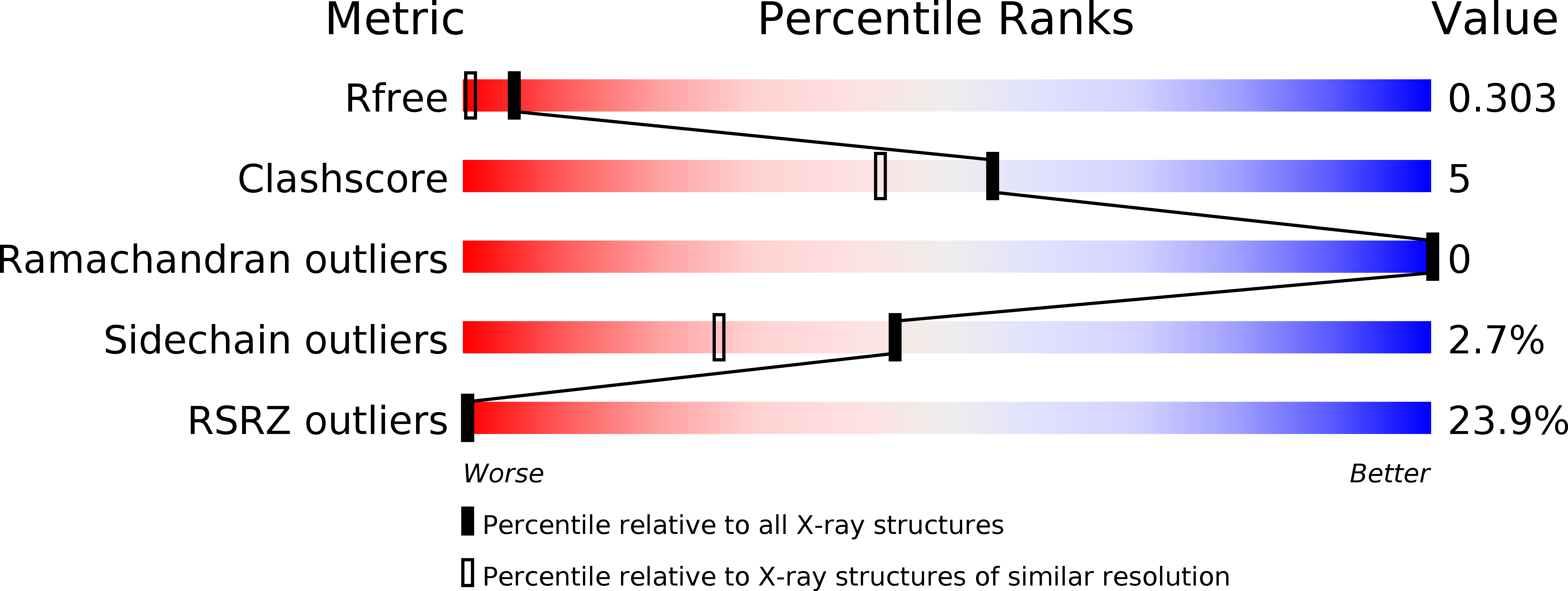
3CG6 image
Deposition Date 2008-03-05
Release Date 2008-04-29
Last Version Date 2024-02-21
Entry Detail
PDB ID:
3CG6
Keywords:
Title:
Crystal structure of Gadd45 gamma
Biological Source:
Source Organism:
Mus musculus (Taxon ID: 10090)
Method Details:
Experimental Method:
Resolution:
1.70 Å
R-Value Free:
0.27
R-Value Work:
0.22
R-Value Observed:
0.22
Space Group:
C 2 2 21
Macromolecular Entities
Structures with similar UniProt ID
Protein Blast
Polymer Type:polypeptide(L)
Molecule:Growth arrest and DNA-damage-inducible 45 gamma
Chain IDs:A, B
Chain Length:146
Number of Molecules:2
Biological Source:Mus musculus
Primary Citation
The crystal structure and dimerization interface of GADD45gamma.
Proc.Natl.Acad.Sci.Usa 105 6566 6571 (2008)
PMID: 18445651 DOI: 10.1073/pnas.0800086105

Abstact

Gadd45 proteins are recognized as tumor and autoimmune suppressors whose expression can be induced by genotoxic stresses. These proteins are involved in cell cycle control, growth arrest, and apoptosis through interactions with a wide variety of binding partners. We report here the crystal structure of Gadd45gamma, which reveals a fold comprising an alphabetaalpha sandwich with a central five-stranded mixed beta-sheet with alpha-helices packed on either side. Based on crystallographic symmetry we identified the dimer interface of Gadd45gamma dimers by generating point mutants that compromised dimerization while leaving the tertiary structure of the monomer intact. The dimer interface comprises a four-helix bundle involving residues that are the most highly conserved among Gadd45 isoforms. Cell-based assays using these point mutants demonstrate that dimerization is essential for growth inhibition. This structural information provides a new context for evaluation of the plethora of protein-protein interactions that govern the many functions of the Gadd45 family of proteins.

Legend

Protein

Chemical

Disease

Primary Citation of related structures