Deposition Date
2008-03-03
Release Date
2009-01-13
Last Version Date
2024-11-20
Entry Detail
PDB ID:
3CFI
Keywords:
Title:
Nanobody-aided structure determination of the EPSI:EPSJ pseudopilin heterdimer from Vibrio Vulnificus
Biological Source:
Source Organism(s):
Vibrio vulnificus (Taxon ID: 672)
Lama glama (Taxon ID: 9844)
Lama glama (Taxon ID: 9844)
Expression System(s):
Method Details:
Experimental Method:
Resolution:
2.58 Å
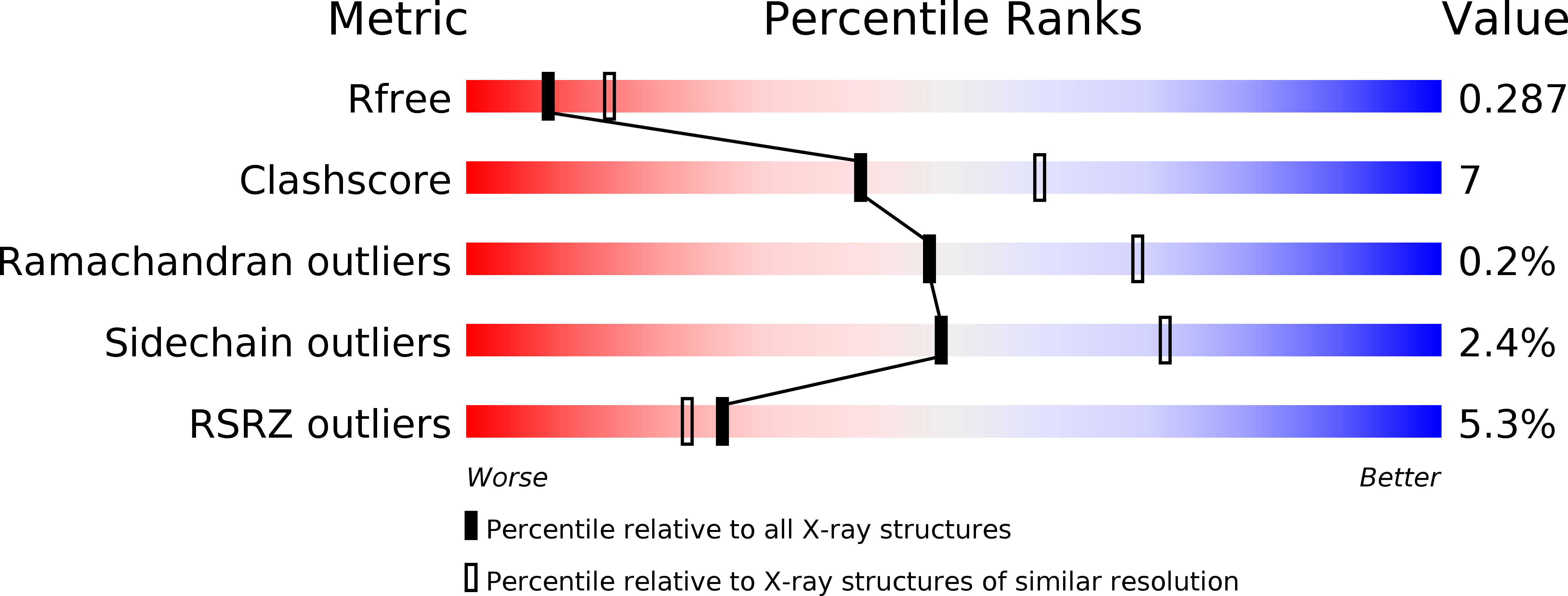
R-Value Free:
0.27
R-Value Work:
0.22
R-Value Observed:
0.23
Space Group:
P 1