Deposition Date
2008-02-29
Release Date
2009-03-03
Last Version Date
2024-02-21
Entry Detail
PDB ID:
3CES
Keywords:
Title:
Crystal Structure of E.coli MnmG (GidA), a Highly-Conserved tRNA Modifying Enzyme
Biological Source:
Source Organism(s):
Escherichia coli (Taxon ID: 562)
Expression System(s):
Method Details:
Experimental Method:
Resolution:
2.41 Å
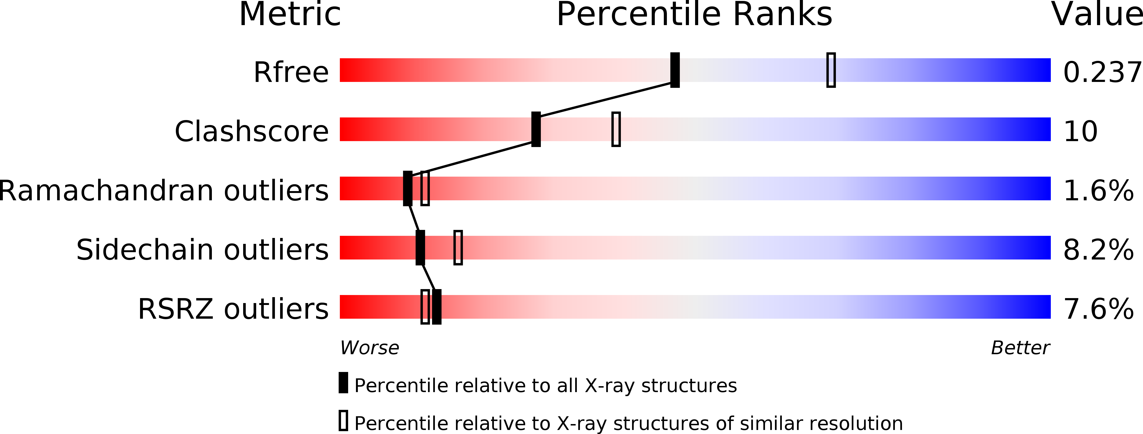
R-Value Free:
0.23
R-Value Work:
0.20
R-Value Observed:
0.20
Space Group:
P 1 21 1