Deposition Date
2008-02-27
Release Date
2008-07-22
Last Version Date
2023-11-01
Entry Detail
PDB ID:
3CDU
Keywords:
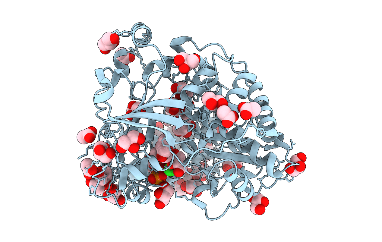
Title:
Crystal structure of coxsackievirus B3 RNA-dependent RNA polymerase (3Dpol) in complex with a pyrophosphate
Biological Source:
Source Organism(s):
Coxsackievirus B3 (Taxon ID: 103903)
Expression System(s):
Method Details:
Experimental Method:
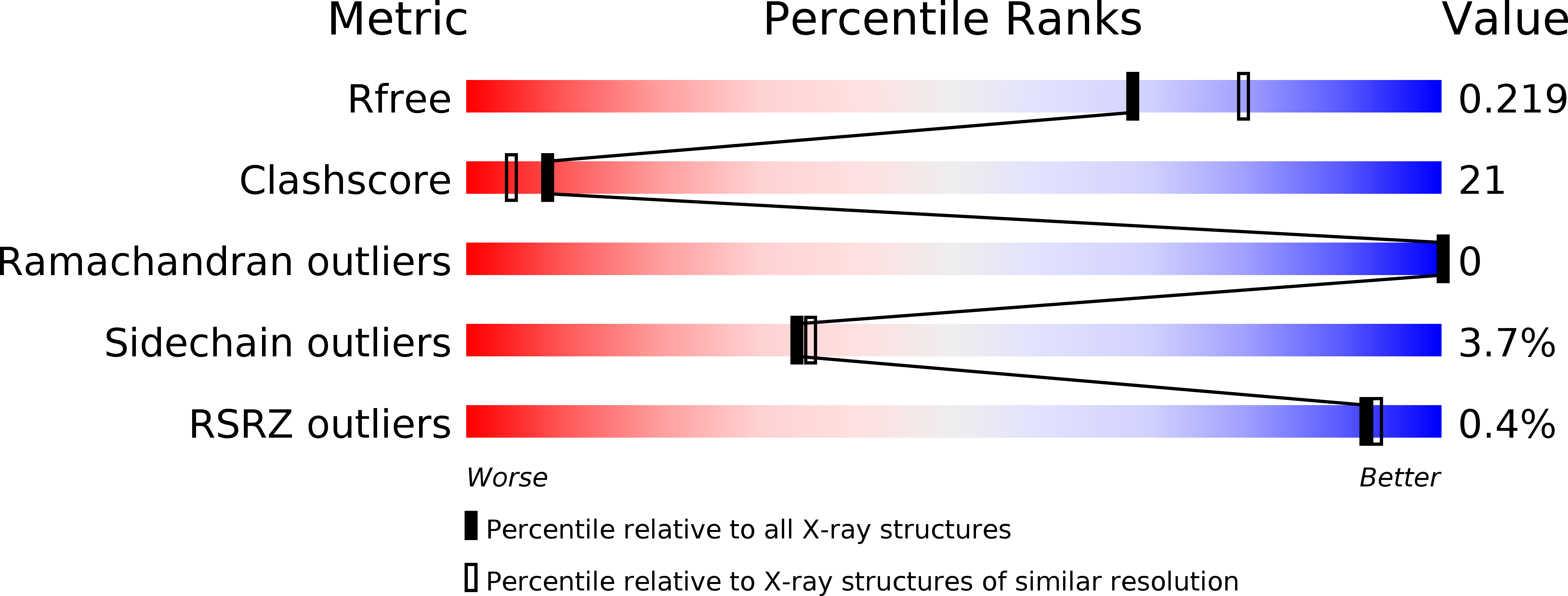
Resolution:
2.10 Å
R-Value Free:
0.21
R-Value Work:
0.17
R-Value Observed:
0.18
Space Group:
P 43 21 2