3CDN image
Deposition Date 2008-02-27
Release Date 2008-06-10
Last Version Date 2024-10-30
Entry Detail
PDB ID:
3CDN
Title:
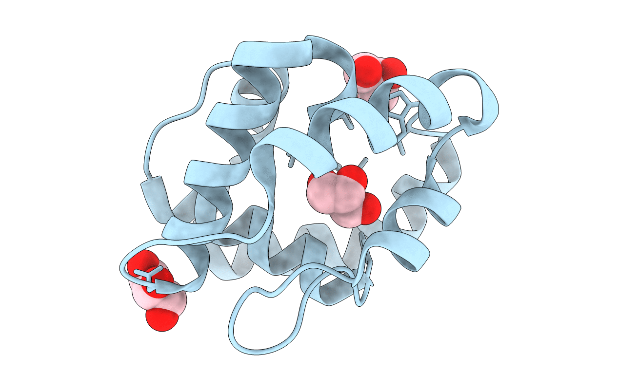
Crystal structure of a pheromone binding protein from Apis mellifera soaked at pH 4.0
Biological Source:
Source Organism:
Apis mellifera (Taxon ID: 7460)
Host Organism:
Method Details:
Experimental Method:
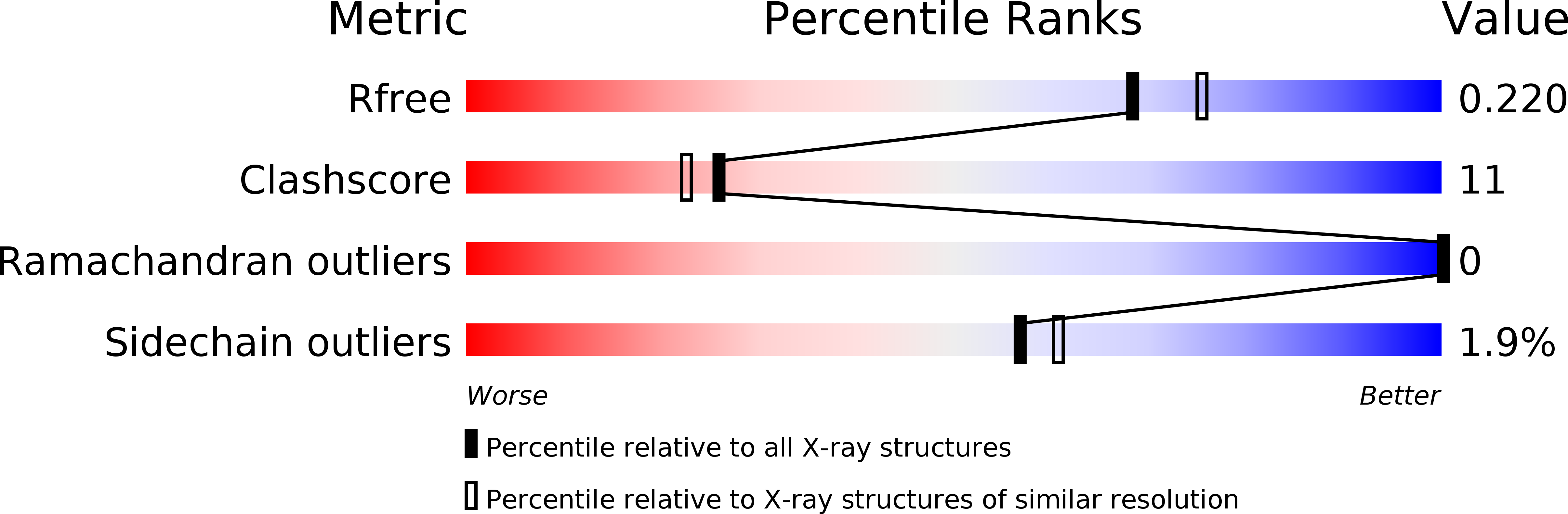
Resolution:
2.00 Å
R-Value Free:
0.20
R-Value Work:
0.17
R-Value Observed:
0.17
Space Group:
C 2 2 21
Macromolecular Entities
Polymer Type:polypeptide(L)
Molecule:Pheromone-binding protein ASP1
Chain IDs:A
Chain Length:119
Number of Molecules:1
Biological Source:Apis mellifera
Primary Citation
Structural basis of the honey bee PBP pheromone and pH-induced conformational change
J.Mol.Biol. 380 158 169 (2008)
PMID: 18508083 DOI: 10.1016/j.jmb.2008.04.048

Abstact

The behavior of insects and their perception of their surroundings are driven, in a large part, by odorants and pheromones. This is especially true for social insects, such as the honey bee, where the queen controls the development and the caste status of the other individuals. Pheromone perception is a complex phenomenon relying on a cascade of recognition events, initiated in antennae by pheromone recognition by a pheromone-binding protein and finishing with signal transduction at the axon membrane level. With to the objective of deciphering this initial step, we have determined the structures of the bee antennal pheromone-binding protein (ASP1) in the apo form and in complex with the main component of the queen mandibular pheromonal mixture, 9-keto-2(E)-decenoic acid (9-ODA) and with nonpheromonal components. In the apo protein, the C terminus obstructs the binding site. In contrast, ASP1 complexes have different open conformations, depending on the ligand shape, leading to different volumes of the binding cavity. The binding site integrity depends on the C terminus (111-119) conformation, which involves the interplay of two factors; i.e. the presence of a ligand and a low pH. Ligand binding to ASP1 is favored by low pH, opposite to what is observed with other pheromone-binding proteins, such as those of Bombyx mori and Anopheles gambiae.

Legend

Protein

Chemical

Disease

Primary Citation of related structures