3CDF image
Deposition Date 2008-02-26
Release Date 2008-09-02
Last Version Date 2024-10-30
Entry Detail
PDB ID:
3CDF
Keywords:
Title:
kI O18/O8 Y87H immunoglobulin light chain variable domain
Biological Source:
Source Organism:
Homo sapiens (Taxon ID: 9606)
Host Organism:
Method Details:
Experimental Method:
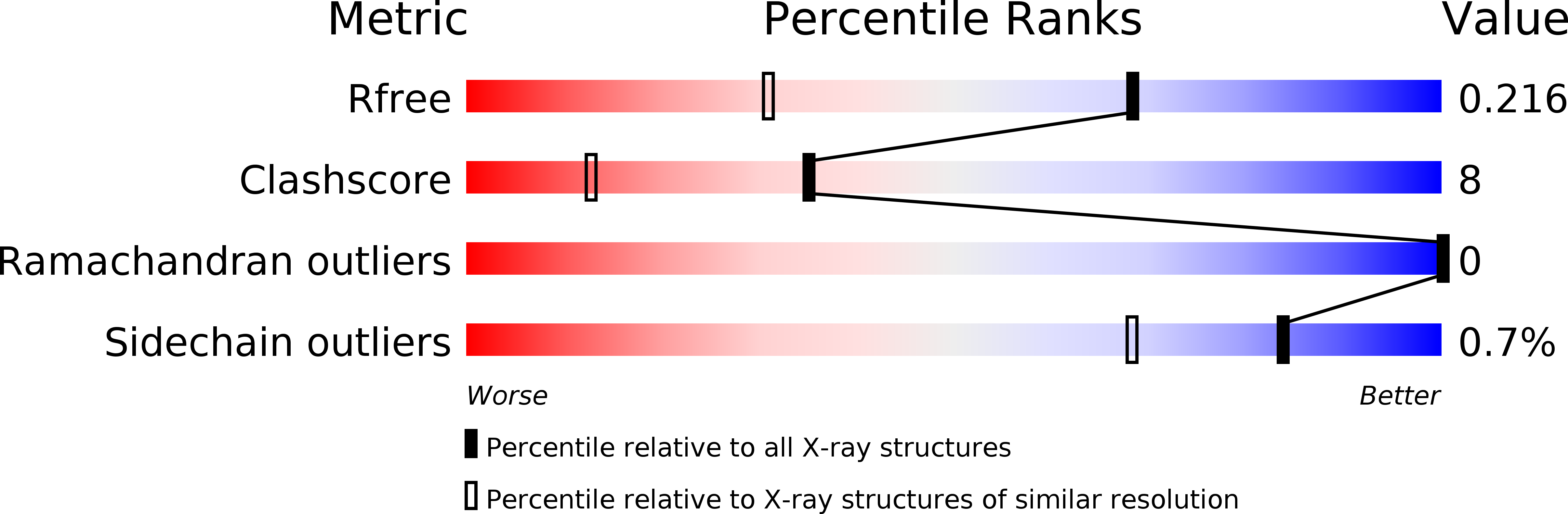
Resolution:
1.53 Å
R-Value Free:
0.23
R-Value Work:
0.18
R-Value Observed:
0.19
Space Group:
P 1 21 1
Macromolecular Entities
Structures with similar UniProt ID
Protein Blast
Polymer Type:polypeptide(L)
Molecule:IMMUNOGLOBULIN LIGHT CHAIN
Mutagens:Y87H
Chain IDs:A, B, C, D, E, F
Chain Length:109
Number of Molecules:6
Biological Source:Homo sapiens
Primary Citation
Structural insights into the role of mutations in amyloidogenesis.
J.Biol.Chem. 283 30950 30956 (2008)
PMID: 18768467 DOI: 10.1074/jbc.M804822200

Abstact

Mechanisms of amyloidogenesis are not well understood, including potential structural contributions of mutations in the process. Our previous research indicated that the dimer interface of amyloidogenic immunoglobulin light chain protein AL-09 is twisted 90 degrees relative to the protein from its germline sequence, kappaI O18/O8. Here we report a systematic restoration of AL-09 to its germline sequence by mutating the non-conservative somatic mutations located in the light chain dimer interface. Among these mutants, we find a correlation between increased thermodynamic stability and an increase in the lag time for fibril formation. The restorative mutant AL-09 H87Y completes the trifecta and restores the dimer interface observed in kappaI O18/O8, emphasizing the potential importance of the structural integrity of these proteins to protect against amyloidogenicity. We also find that adding amyloidogenic mutations into the germline protein illustrates mutational cooperativity in promoting amyloidogenesis.

Legend

Protein

Chemical

Disease

Primary Citation of related structures