3CAP image
Entry Detail
PDB ID:
3CAP
Title:
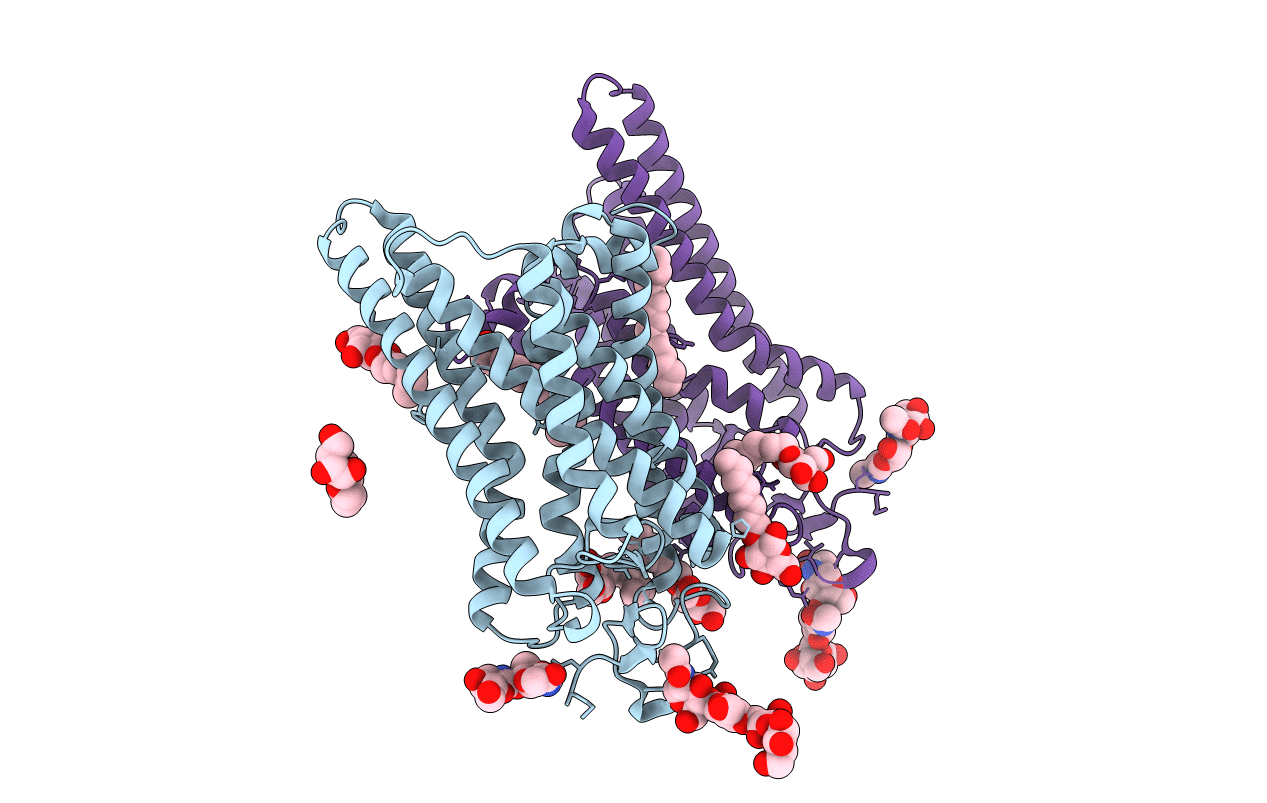
Crystal Structure of Native Opsin: the G Protein-Coupled Receptor Rhodopsin in its Ligand-free State
Biological Source:
Source Organism:
PDB Version:
Deposition Date:
2008-02-20
Release Date:
2008-06-24
Method Details:
Experimental Method:
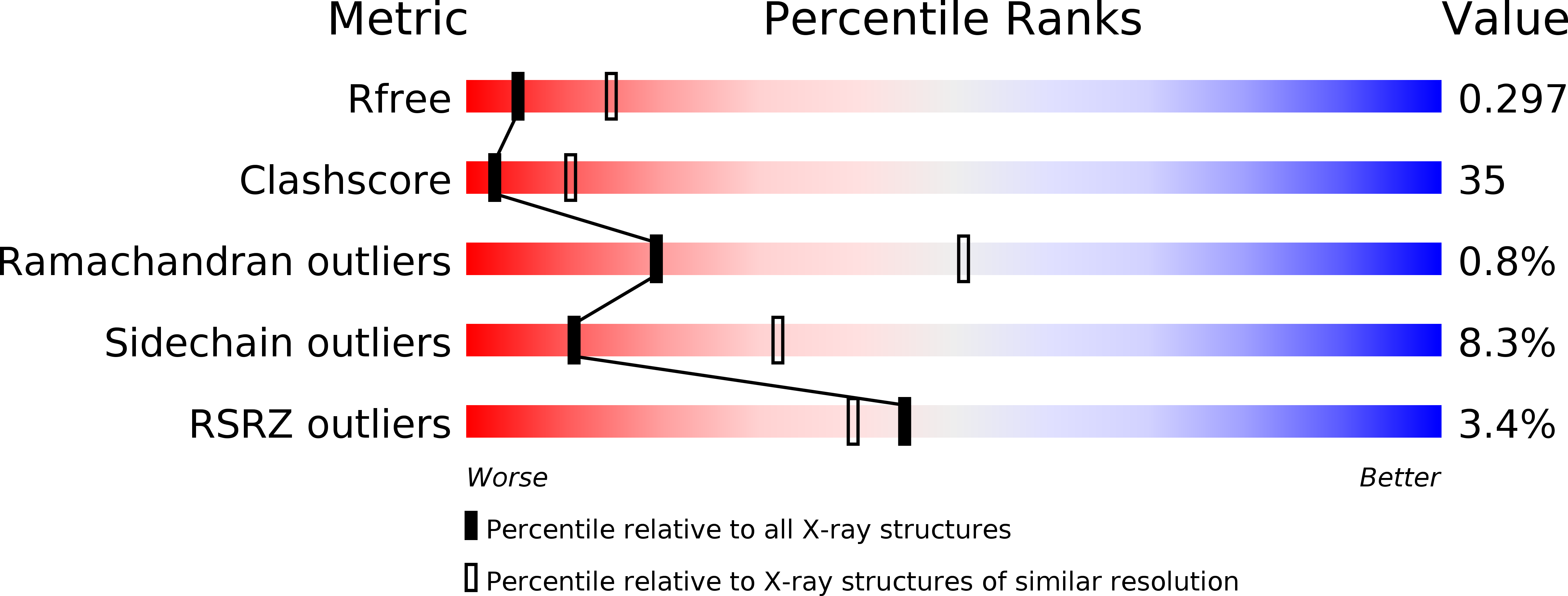
Resolution:
2.90 Å
R-Value Free:
0.26
R-Value Work:
0.22
R-Value Observed:
0.23
Space Group:
H 3
Macromolecular Entities
Polymer Type:polypeptide(L)
Description:Rhodopsin
Chain IDs:A, B
Chain Length:348
Number of Molecules:2
Biological Source:Bos taurus
Modified Residue
Compound ID Chain ID Parent Comp ID Details 2D Image
ASN A ASN GLYCOSYLATION SITE
Primary Citation
Crystal structure of the ligand-free G-protein-coupled receptor opsin
Nature 454 183 187 (2008)
PMID: 18563085 DOI: 10.1038/nature07063

Abstact

In the G-protein-coupled receptor (GPCR) rhodopsin, the inactivating ligand 11-cis-retinal is bound in the seven-transmembrane helix (TM) bundle and is cis/trans isomerized by light to form active metarhodopsin II. With metarhodopsin II decay, all-trans-retinal is released, and opsin is reloaded with new 11-cis-retinal. Here we present the crystal structure of ligand-free native opsin from bovine retinal rod cells at 2.9 ångström (A) resolution. Compared to rhodopsin, opsin shows prominent structural changes in the conserved E(D)RY and NPxxY(x)(5,6)F regions and in TM5-TM7. At the cytoplasmic side, TM6 is tilted outwards by 6-7 A, whereas the helix structure of TM5 is more elongated and close to TM6. These structural changes, some of which were attributed to an active GPCR state, reorganize the empty retinal-binding pocket to disclose two openings that may serve the entry and exit of retinal. The opsin structure sheds new light on ligand binding to GPCRs and on GPCR activation.

Legend

Protein

Chemical

Disease

Primary Citation of related structures