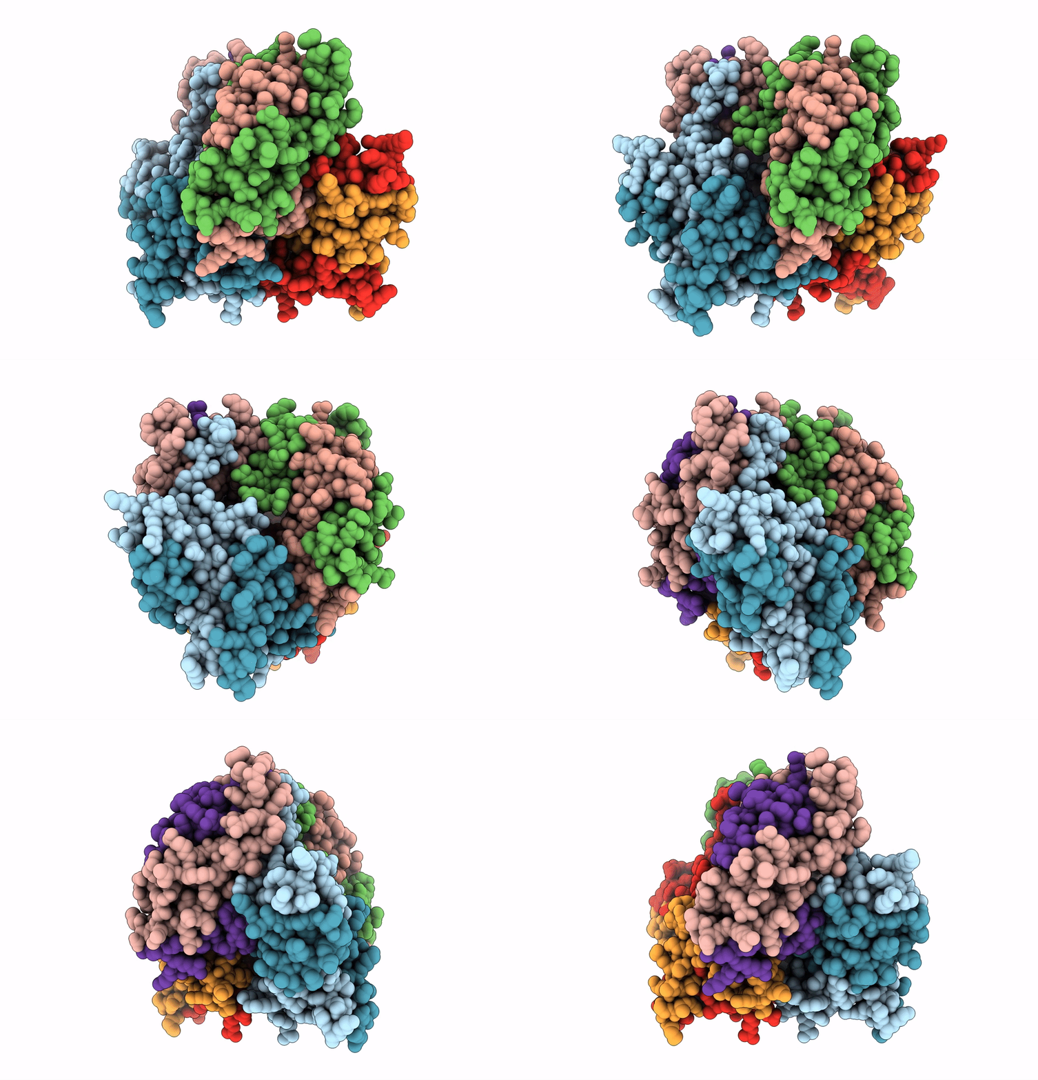
3C9K image
Deposition Date 2008-02-16
Release Date 2009-01-06
Last Version Date 2024-03-13
Entry Detail
PDB ID:
3C9K
Title:
Model of Histone Octamer Tubular Crystals
Biological Source:
Source Organism:
Gallus gallus (Taxon ID: 9031)
Method Details:
Experimental Method:
Resolution:
20.00 Å
Aggregation State:
FILAMENT
Reconstruction Method:
HELICAL
Macromolecular Entities
Structures with similar UniProt ID
Protein Blast
Polymer Type:polypeptide(L)
Molecule:Histone H2A-IV
Chain IDs:A, E
Chain Length:128
Number of Molecules:2
Biological Source:Gallus gallus
Structures with similar UniProt ID
Protein Blast
Polymer Type:polypeptide(L)
Molecule:Histone H2B 7
Gene (Uniprot):H2B-VII
Chain IDs:B, F
Chain Length:125
Number of Molecules:2
Biological Source:Gallus gallus
Structures with similar UniProt ID
Protein Blast
Polymer Type:polypeptide(L)
Molecule:Histone H3.2
Gene (Uniprot):H3-I, H3-II, H3-III, H3-IV, H3-V, H3-VI, H3-VII, H3-VIII
Chain IDs:C, G
Chain Length:135
Number of Molecules:2
Biological Source:Gallus gallus
Structures with similar UniProt ID
Protein Blast
Polymer Type:polypeptide(L)
Molecule:Histone H4
Gene (Uniprot):H4-I, H4-II, H4-III, H4-IV, H4-V, H4-VI, H4-VII
Chain IDs:D, H
Chain Length:102
Number of Molecules:2
Biological Source:Gallus gallus
Ligand Molecules
Primary Citation
Histone octamer helical tubes suggest that an internucleosomal four-helix bundle stabilizes the chromatin fiber
Biophys.J. 96 3363 3371 (2009)
PMID: 19383479 DOI: 10.1016/j.bpj.2008.10.075

Abstact

A major question in chromatin involves the exact organization of nucleosomes within the 30-nm chromatin fiber and its structural determinants of assembly. Here we investigate the structure of histone octamer helical tubes via the method of iterative helical real-space reconstruction. Accurate placement of the x-ray structure of the histone octamer within the reconstructed density yields a pseudoatomic model for the entire helix, and allows precise identification of molecular interactions between neighboring octamers. One such interaction that would not be obscured by DNA in the nucleosome consists of a twofold symmetric four-helix bundle formed between pairs of H2B-alpha3 and H2B-alphaC helices of neighboring octamers. We believe that this interface can act as an internucleosomal four-helix bundle within the context of the chromatin fiber. The potential relevance of this interface in the folding of the 30-nm chromatin fiber is discussed.

Legend

Protein

Chemical

Disease

Primary Citation of related structures