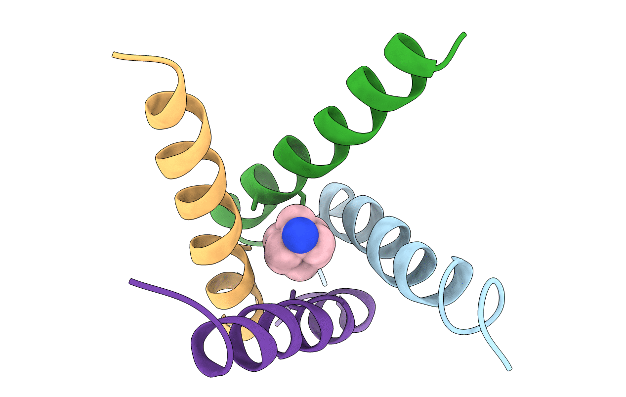
3C9J image
Deposition Date 2008-02-15
Release Date 2008-03-11
Last Version Date 2024-04-03
Entry Detail
PDB ID:
3C9J
Title:
The Crystal structure of Transmembrane domain of M2 protein and Amantadine complex
Biological Source:
Source Organism:
(Taxon ID: )
Method Details:
Experimental Method:
Resolution:
3.50 Å
R-Value Free:
0.31
R-Value Work:
0.29
R-Value Observed:
0.29
Space Group:
P 21 21 2
Macromolecular Entities
Polymer Type:polypeptide(L)
Molecule:Proton Channel protein M2, transmembrane segment
Gene (Uniprot):M
Mutations:G34A
Chain IDs:A, B, C, D
Chain Length:25
Number of Molecules:4
Biological Source:
Ligand Molecules
Primary Citation
Structural basis for the function and inhibition of an influenza virus proton channel
Nature 451 596 600 (2008)
PMID: 18235504 DOI: 10.1038/nature06528

Abstact

The M2 protein from influenza A virus is a pH-activated proton channel that mediates acidification of the interior of viral particles entrapped in endosomes. M2 is the target of the anti-influenza drugs amantadine and rimantadine; recently, resistance to these drugs in humans, birds and pigs has reached more than 90% (ref. 1). Here we describe the crystal structure of the transmembrane-spanning region of the homotetrameric protein in the presence and absence of the channel-blocking drug amantadine. pH-dependent structural changes occur near a set of conserved His and Trp residues that are involved in proton gating. The drug-binding site is lined by residues that are mutated in amantadine-resistant viruses. Binding of amantadine physically occludes the pore, and might also perturb the pK(a) of the critical His residue. The structure provides a starting point for solving the problem of resistance to M2-channel blockers.

Legend

Protein

Chemical

Disease

Primary Citation of related structures