Deposition Date
2008-02-15
Release Date
2008-05-13
Last Version Date
2023-08-30
Entry Detail
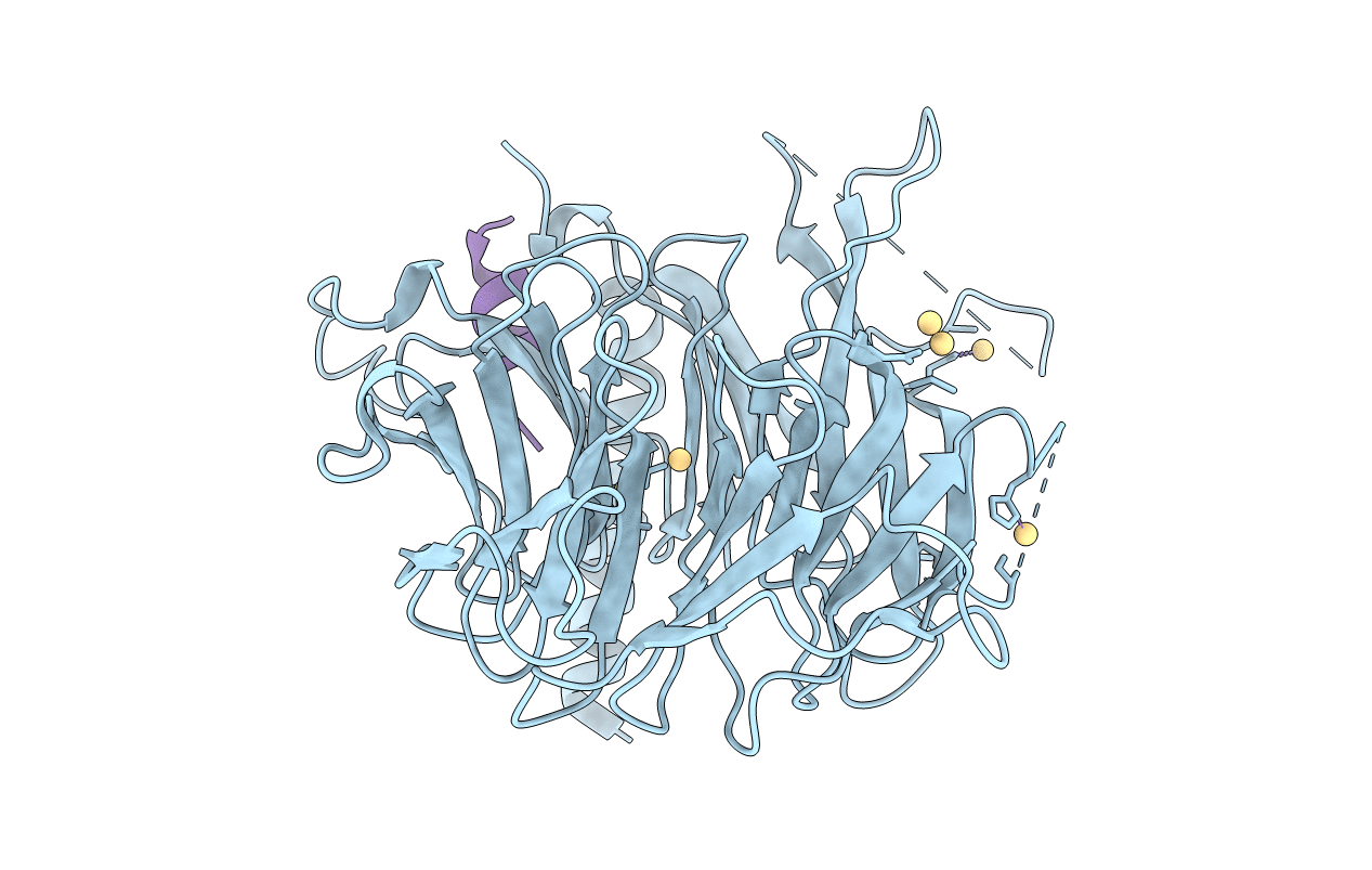
PDB ID:
3C9C
Keywords:
Title:
Structural Basis of Histone H4 Recognition by p55
Biological Source:
Source Organism(s):
Drosophila melanogaster (Taxon ID: 7227)
Expression System(s):
Method Details:
Experimental Method:
Resolution:
3.20 Å
R-Value Free:
0.24
R-Value Work:
0.20
Space Group:
P 31 2 1