
Deposition Date
2008-02-11
Release Date
2008-04-22
Last Version Date
2024-10-30
Entry Detail
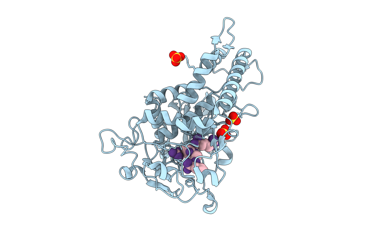
PDB ID:
3C8A
Keywords:
Title:
Crystal structure of the catalytic domain of botulinum neurotoxin serotype A with inhibitory peptide RRGL
Biological Source:
Source Organism(s):
Clostridium botulinum (Taxon ID: 1491)
synthetic construct (Taxon ID: 32630)
synthetic construct (Taxon ID: 32630)
Expression System(s):
Method Details:
Experimental Method:
Resolution:
1.52 Å
R-Value Free:
0.20
R-Value Work:
0.19
R-Value Observed:
0.19
Space Group:
P 1 21 1
