
Deposition Date
2008-02-07
Release Date
2008-05-06
Last Version Date
2023-08-30
Entry Detail
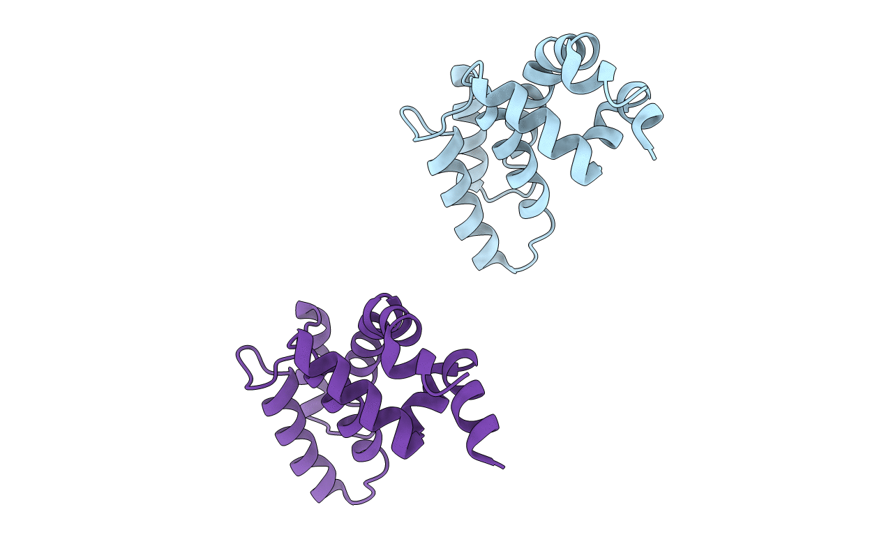
PDB ID:
3C7L
Keywords:
Title:
Molecular architecture of Galphao and the structural basis for RGS16-mediated deactivation
Biological Source:
Source Organism(s):
Mus musculus (Taxon ID: 10090)
Expression System(s):
Method Details:
Experimental Method:
Resolution:
1.89 Å
R-Value Free:
0.26
R-Value Work:
0.22
Space Group:
P 21 21 21
