3C75 image
Deposition Date 2008-02-06
Release Date 2008-12-30
Last Version Date 2025-03-26
Entry Detail
PDB ID:
3C75
Keywords:
Title:
Paracoccus versutus methylamine dehydrogenase in complex with amicyanin
Biological Source:
Source Organism(s):
Method Details:
Experimental Method:
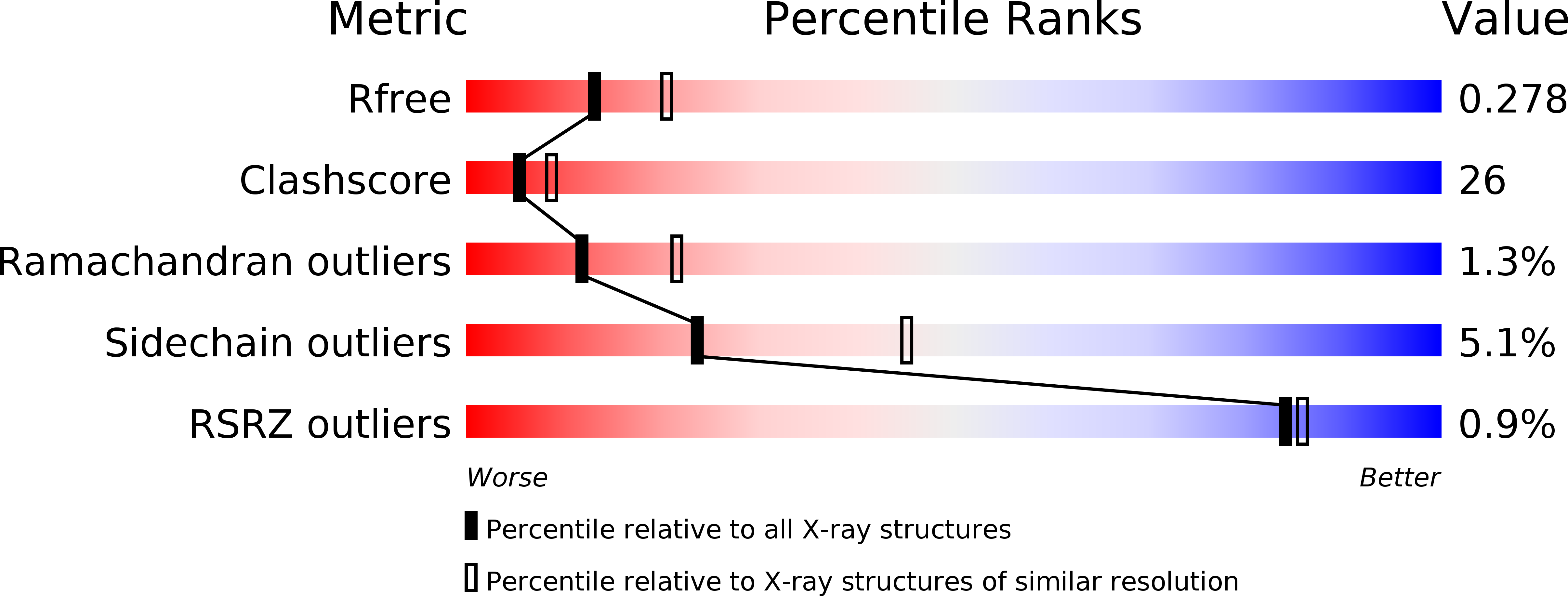
Resolution:
2.50 Å
R-Value Free:
0.28
R-Value Work:
0.23
R-Value Observed:
0.23
Space Group:
P 21 21 21
Macromolecular Entities
Structures with similar UniProt ID
Protein Blast
Polymer Type:polypeptide(L)
Molecule:Amicyanin
Gene (Uniprot):mauC
Chain IDs:C (auth: A), F (auth: B)
Chain Length:132
Number of Molecules:2
Biological Source:Paracoccus versutus
Structures with similar UniProt ID
Protein Blast
Polymer Type:polypeptide(L)
Molecule:Methylamine dehydrogenase heavy chain
Gene (Uniprot):mauB
Chain IDs:A (auth: H), D (auth: J)
Chain Length:426
Number of Molecules:2
Biological Source:Paracoccus versutus
Structures with similar UniProt ID
Protein Blast
Polymer Type:polypeptide(L)
Molecule:Methylamine dehydrogenase light chain
Gene (Uniprot):mauA
Chain IDs:B (auth: L), E (auth: M)
Chain Length:188
Number of Molecules:2
Biological Source:Paracoccus versutus
Modified Residue
Compound ID Chain ID Parent Comp ID Details 2D Image
TRQ B TRP ?
Ligand Molecules
Primary Citation
Structural comparison of crystal and solution states of the 138 kDa complex of methylamine dehydrogenase and amicyanin from Paracoccus versutus.
Biochemistry 47 6560 6570 (2008)
PMID: 18512962 DOI: 10.1021/bi7023749

Abstact

Methylamine can be used as the sole carbon source of certain methylotrophic bacteria. Methylamine dehydrogenase catalyzes the conversion of methylamine into formaldehyde and donates electrons to the electron transfer protein amicyanin. The crystal structure of the complex of methylamine dehydrogenase and amicyanin from Paracoccus versutus has been determined, and the rate of electron transfer from the tryptophan tryptophylquinone cofactor of methylamine dehydrogenase to the copper ion of amicyanin in solution has been determined. In the presence of monovalent ions, the rate of electron transfer from the methylamine-reduced TTQ is much higher than in their absence. In general, the kinetics are similar to those observed for the system from Paracoccus denitrificans. The complex in solution has been studied using nuclear magnetic resonance. Signals of perdeuterated, (15)N-enriched amicyanin bound to methylamine dehydrogenase are observed. Chemical shift perturbation analysis indicates that the dissociation rate constant is approximately 250 s(-1) and that amicyanin assumes a well-defined position in the complex in solution. The most affected residues are in the interface observed in the crystal structure, whereas smaller chemical shift changes extend to deep inside the protein. These perturbations can be correlated to small differences in the hydrogen bond network observed in the crystal structures of free and bound amicyanin. This study indicates that chemical shift changes can be used as reliable indicators of subtle structural changes even in a complex larger than 100 kDa.

Legend

Protein

Chemical

Disease

Primary Citation of related structures
Feedback Form
Name
Email
Institute
Feedback