3C50 image
Deposition Date 2008-01-30
Release Date 2008-03-11
Last Version Date 2023-08-30
Entry Detail
PDB ID:
3C50
Keywords:
Title:
Crystal Structure of G protein coupled receptor kinase 1 bound to ADP and magnesium chloride at 2.6A
Biological Source:
Source Organism:
Bos taurus (Taxon ID: 9913)
Host Organism:
Method Details:
Experimental Method:
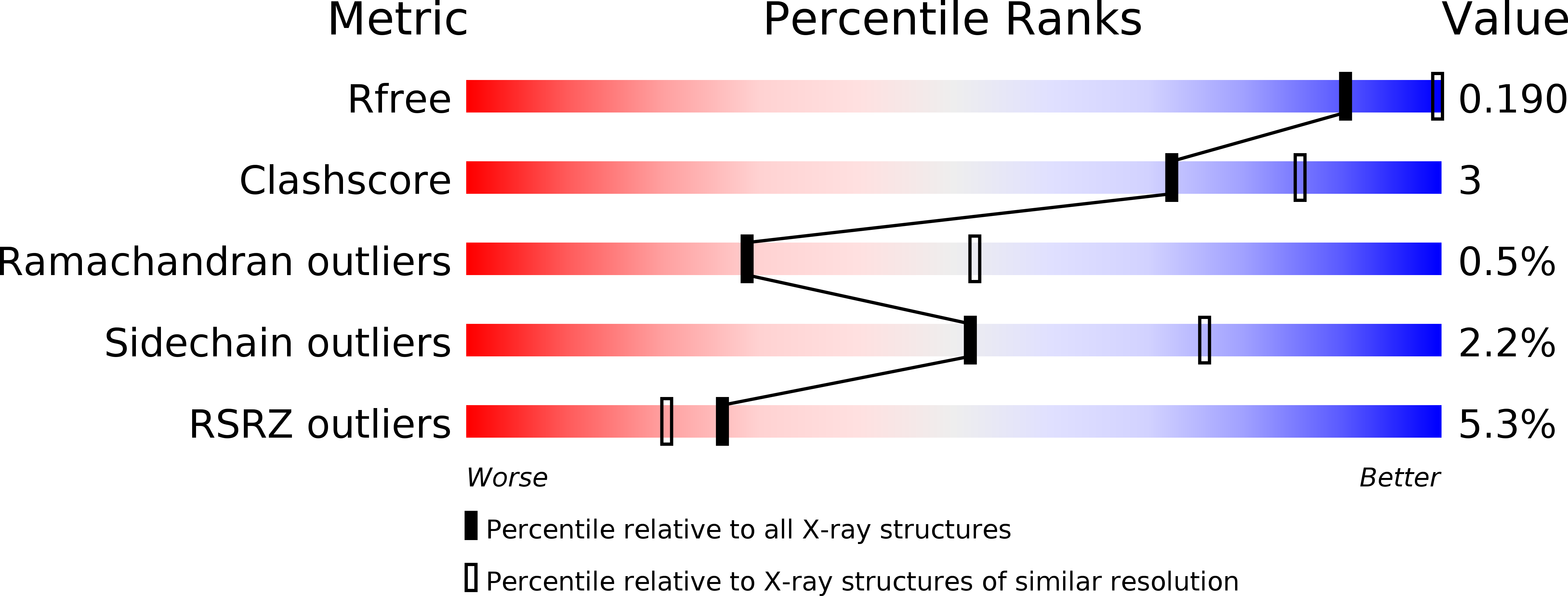
Resolution:
2.60 Å
R-Value Work:
0.18
R-Value Observed:
0.18
Space Group:
P 21 21 21
Macromolecular Entities
Structures with similar UniProt ID
Protein Blast
Polymer Type:polypeptide(L)
Molecule:Rhodopsin kinase
Gene (Uniprot):GRK1
Chain IDs:A, B
Chain Length:543
Number of Molecules:2
Biological Source:Bos taurus
Primary Citation
Structures of rhodopsin kinase in different ligand states reveal key elements involved in G protein-coupled receptor kinase activation.
J.Biol.Chem. 283 14053 14062 (2008)
PMID: 18339619 DOI: 10.1074/jbc.M708974200

Abstact

G protein-coupled receptor (GPCR) kinases (GRKs) phosphorylate activated heptahelical receptors, leading to their uncoupling from G proteins. Here we report six crystal structures of rhodopsin kinase (GRK1), revealing not only three distinct nucleotide-binding states of a GRK but also two key structural elements believed to be involved in the recognition of activated GPCRs. The first is the C-terminal extension of the kinase domain, which was observed in all nucleotide-bound GRK1 structures. The second is residues 5-30 of the N terminus, observed in one of the GRK1.(Mg2+)2.ATP structures. The N terminus was also clearly phosphorylated, leading to the identification of two novel phosphorylation sites by mass spectral analysis. Co-localization of the N terminus and the C-terminal extension near the hinge of the kinase domain suggests that activated GPCRs stimulate kinase activity by binding to this region to facilitate full closure of the kinase domain.

Legend

Protein

Chemical

Disease

Primary Citation of related structures
Feedback Form
Name
Email
Institute
Feedback