3C4P image
Deposition Date 2008-01-30
Release Date 2008-05-27
Last Version Date 2024-11-20
Entry Detail
PDB ID:
3C4P
Title:
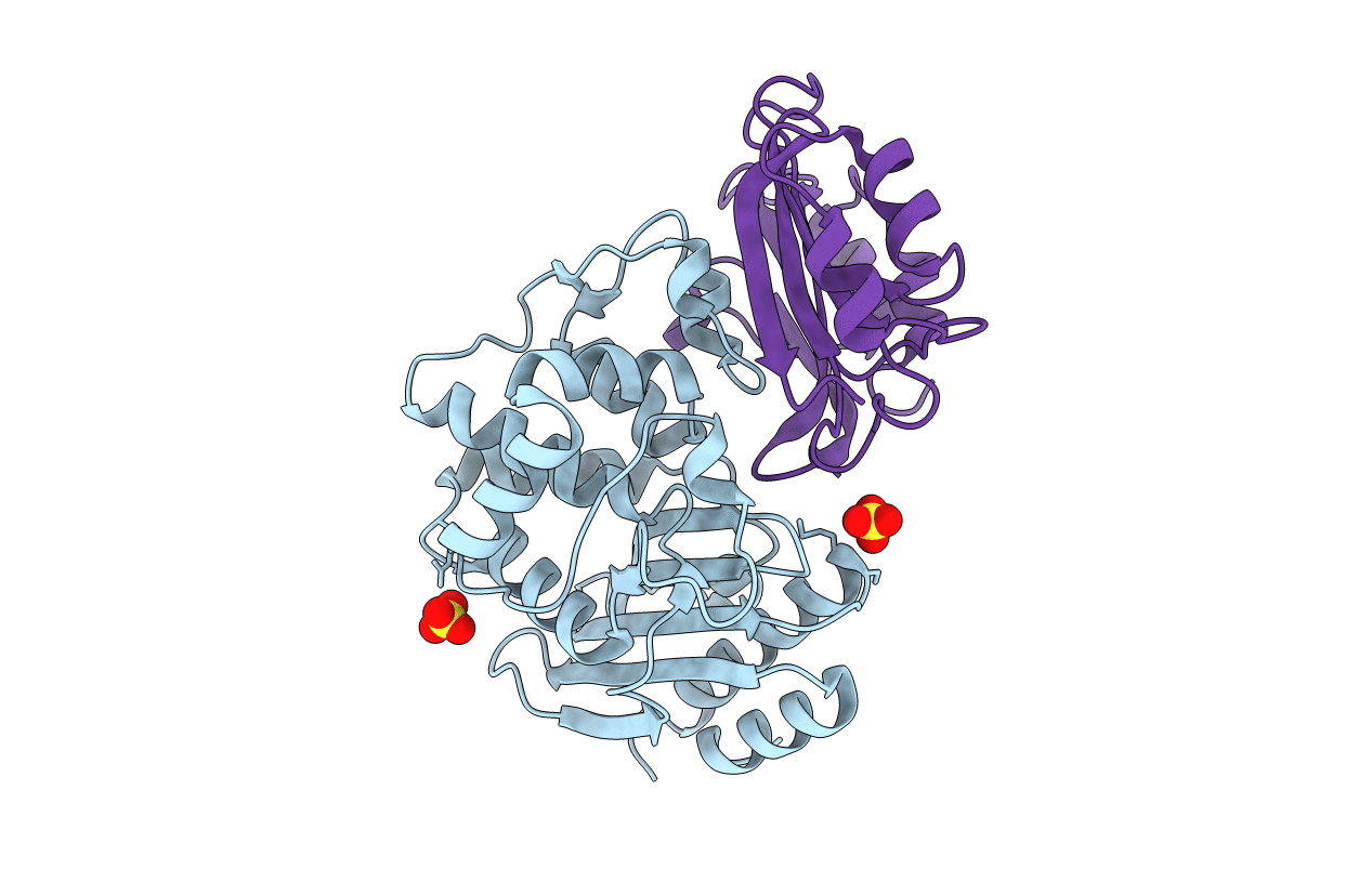
Crystal Structure of the SHV-1 Beta-lactamase/Beta-lactamase inhibitor protein (BLIP) E73M complex
Biological Source:
Source Organism:
Host Organism:
Method Details:
Experimental Method:
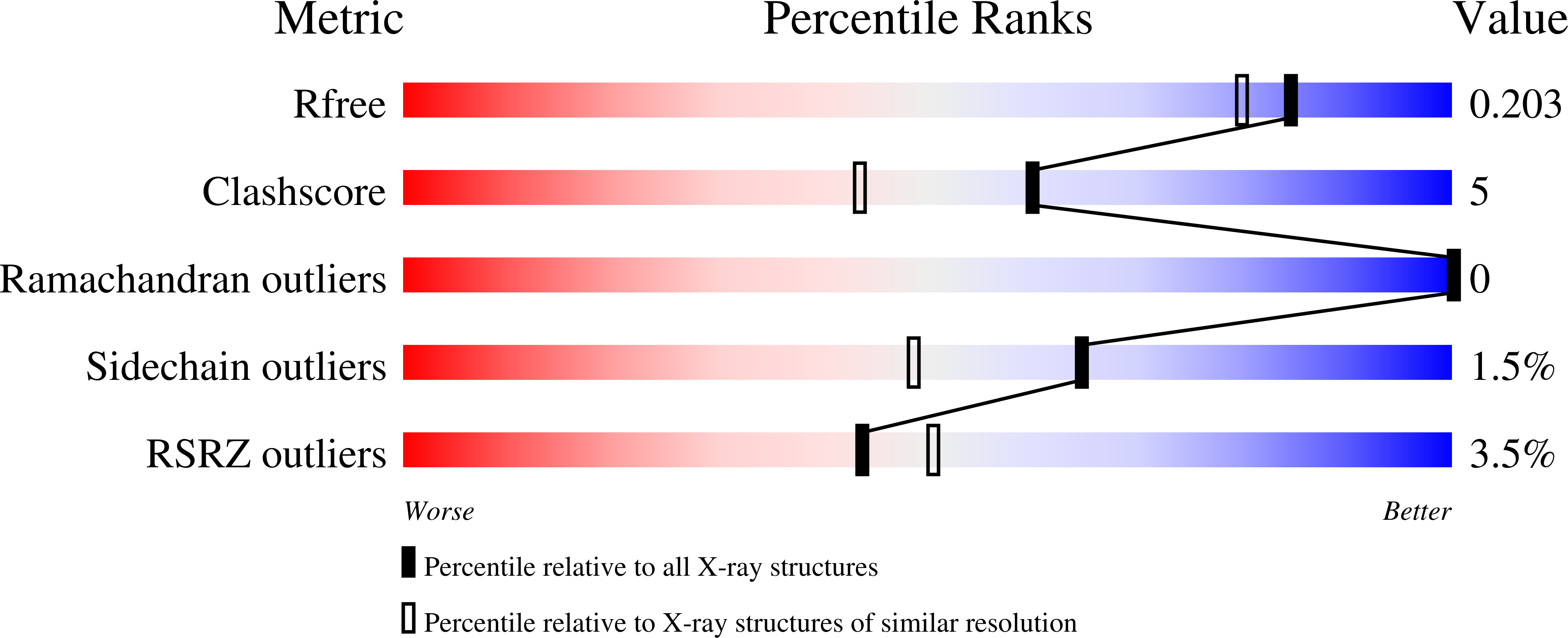
Resolution:
1.75 Å
R-Value Free:
0.20
R-Value Work:
0.18
R-Value Observed:
0.18
Space Group:
P 63
Macromolecular Entities
Polymer Type:polypeptide(L)
Molecule:Beta-lactamase SHV-1
Gene (Uniprot):bla
Chain IDs:A
Chain Length:265
Number of Molecules:1
Biological Source:Klebsiella pneumoniae
Polymer Type:polypeptide(L)
Molecule:Beta-lactamase inhibitory protein
Mutations:E109M
Chain IDs:B
Chain Length:165
Number of Molecules:1
Biological Source:Streptomyces clavuligerus
Ligand Molecules
Primary Citation
Computational redesign of the SHV-1 beta-lactamase/beta-lactamase inhibitor protein interface.
J.Mol.Biol. 382 1265 1275 (2008)
PMID: 18775544 DOI: 10.1016/j.jmb.2008.05.051

Abstact

Beta-lactamases are enzymes that catalyze the hydrolysis of beta-lactam antibiotics. beta-lactamase/beta-lactamase inhibitor protein (BLIP) complexes are emerging as a well characterized experimental model system for studying protein-protein interactions. BLIP is a 165 amino acid protein that inhibits several class A beta-lactamases with a wide range of affinities: picomolar affinity for K1; nanomolar affinity for TEM-1, SME-1, and BlaI; but only micromolar affinity for SHV-1 beta-lactamase. The large differences in affinity coupled with the availability of extensive mutagenesis data and high-resolution crystal structures for the TEM-1/BLIP and SHV-1/BLIP complexes make them attractive systems for the further development of computational design methodology. We used EGAD, a physics-based computational design program, to redesign BLIP in an attempt to increase affinity for SHV-1. Characterization of several of designs and point mutants revealed that in all cases, the mutations stabilize the interface by 10- to 1000-fold relative to wild type BLIP. The calculated changes in binding affinity for the mutants were within a mean absolute error of 0.87 kcal/mol from the experimental values, and comparison of the calculated and experimental values for a set of 30 SHV-1/BLIP complexes yielded a correlation coefficient of 0.77. Structures of the two complexes with the highest affinity, SHV-1/BLIP (E73M) and SHV-1/BLIP (E73M, S130K, S146M), are presented at 1.7 A resolution. While the predicted structures have much in common with the experimentally determined structures, they do not coincide perfectly; in particular a salt bridge between SHV-1 D104 and BLIP K74 is observed in the experimental structures, but not in the predicted design conformations. This discrepancy highlights the difficulty of modeling salt bridge interactions with a protein design algorithm that approximates side chains as discrete rotamers. Nevertheless, while local structural features of the interface were sometimes miscalculated, EGAD is globally successful in designing complexes with increased affinity.

Legend

Protein

Chemical

Disease

Primary Citation of related structures