3C3E image
Deposition Date 2008-01-28
Release Date 2008-02-19
Last Version Date 2024-10-16
Entry Detail
PDB ID:
3C3E
Keywords:
Title:
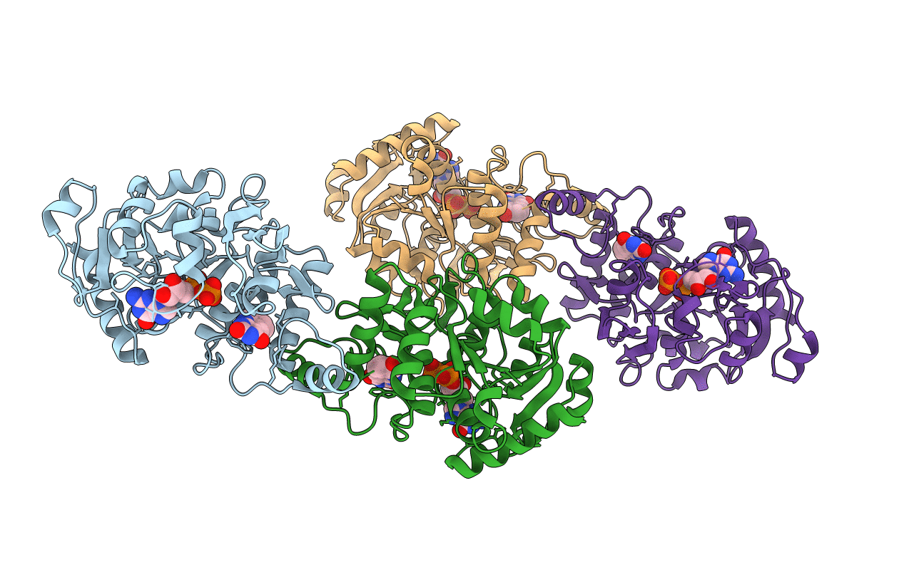
Crystal structure of 2-phospho-(S)-lactate transferase from Methanosarcina mazei in complex with Fo and GDP. Northeast Structural Genomics Consortium target MaR46
Biological Source:
Source Organism:
Host Organism:
Method Details:
Experimental Method:
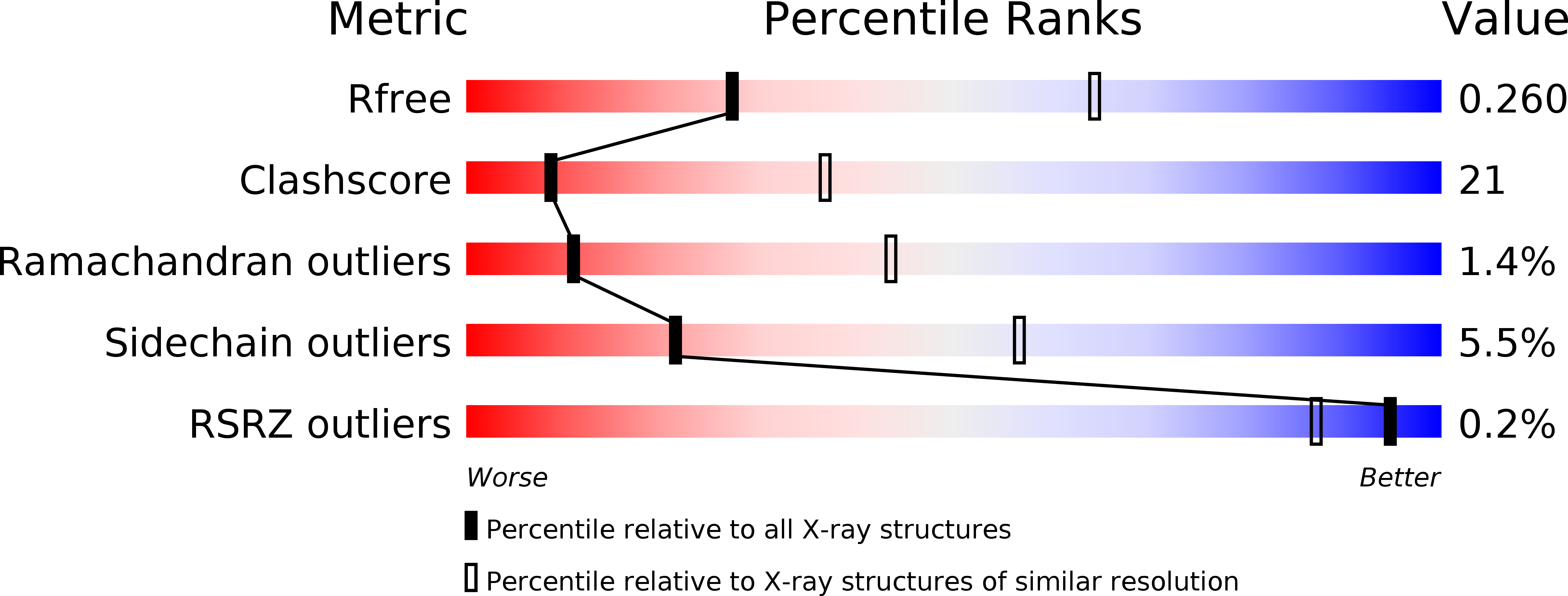
Resolution:
3.00 Å
R-Value Free:
0.23
R-Value Work:
0.20
R-Value Observed:
0.20
Space Group:
P 32
Macromolecular Entities
Polymer Type:polypeptide(L)
Molecule:2-phospho-L-lactate transferase
Gene (Uniprot):cofD
Chain IDs:A, B, C, D
Chain Length:311
Number of Molecules:4
Biological Source:Methanosarcina mazei Go1
Modified Residue
Compound ID Chain ID Parent Comp ID Details 2D Image
MSE A MET SELENOMETHIONINE
Primary Citation
Molecular insights into the biosynthesis of the f420 coenzyme.
J.Biol.Chem. 283 11832 11840 (2008)
PMID: 18252724 DOI: 10.1074/jbc.M710352200

Abstact

Coenzyme F(420), a hydride carrier, is found in Archaea and some bacteria and has crucial roles in methanogenesis, antibiotic biosynthesis, DNA repair, and activation of antitubercular compounds. CofD, 2-phospho-l-lactate transferase, catalyzes the last step in the biosynthesis of F(420)-0 (F(420) without polyglutamate), by transferring the lactyl phosphate moiety of lactyl(2)diphospho-(5')guanosine to 7,8-didemethyl-8-hydroxy-5-deazariboflavin ribitol (Fo). CofD is highly conserved among F(420)-producing organisms, and weak sequence homologs are also found in non-F(420)-producing organisms. This superfamily does not share any recognizable sequence conservation with other proteins. Here we report the first crystal structures of CofD, the free enzyme and two ternary complexes, with Fo and P(i) or with Fo and GDP, from Methanosarcina mazei. The active site is located at the C-terminal end of a Rossmann fold core, and three large insertions make significant contributions to the active site and dimer formation. The observed binding modes of Fo and GDP can explain known biochemical properties of CofD and are also supported by our binding assays. The structures provide significant molecular insights into the biosynthesis of the F(420) coenzyme. Large structural differences in the active site region of the non-F(420)-producing CofD homologs suggest that they catalyze a different biochemical reaction.

Legend

Protein

Chemical

Disease

Primary Citation of related structures