Deposition Date
2008-01-25
Release Date
2008-12-09
Last Version Date
2024-02-21
Entry Detail
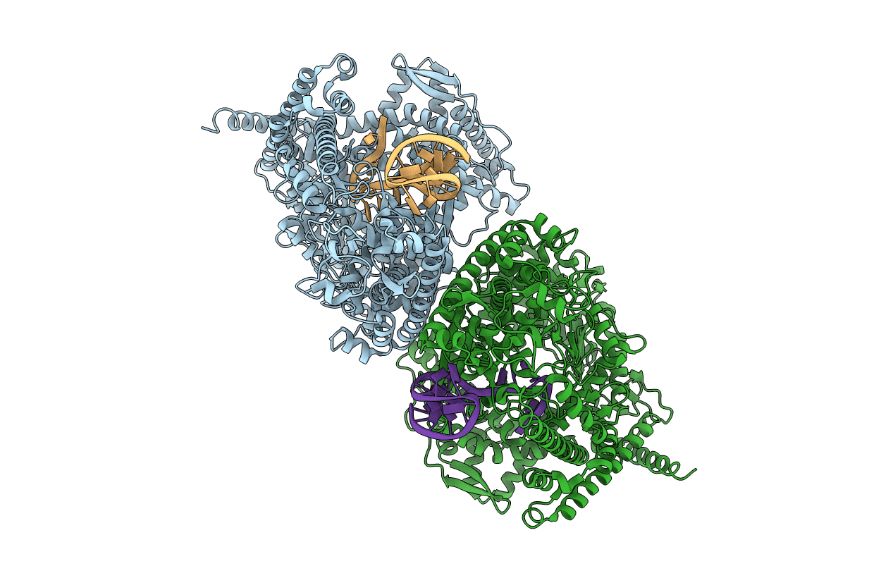
PDB ID:
3C2P
Keywords:
Title:
X-ray crystal structure of the N4 mini-vRNAP P1 promoter complex
Biological Source:
Source Organism(s):
Bacteriophage N4 (Taxon ID: )
Expression System(s):
Method Details:
Experimental Method:
Resolution:
2.00 Å
R-Value Free:
0.25
R-Value Work:
0.22
Space Group:
P 21 21 21