Deposition Date
2008-01-25
Release Date
2008-06-03
Last Version Date
2024-03-13
Entry Detail
PDB ID:
3C2J
Keywords:
Title:
Crystal structure analysis of trioxacarcin A covalently bound to d(AACCGGTT)
Method Details:
Experimental Method:
Resolution:
1.78 Å
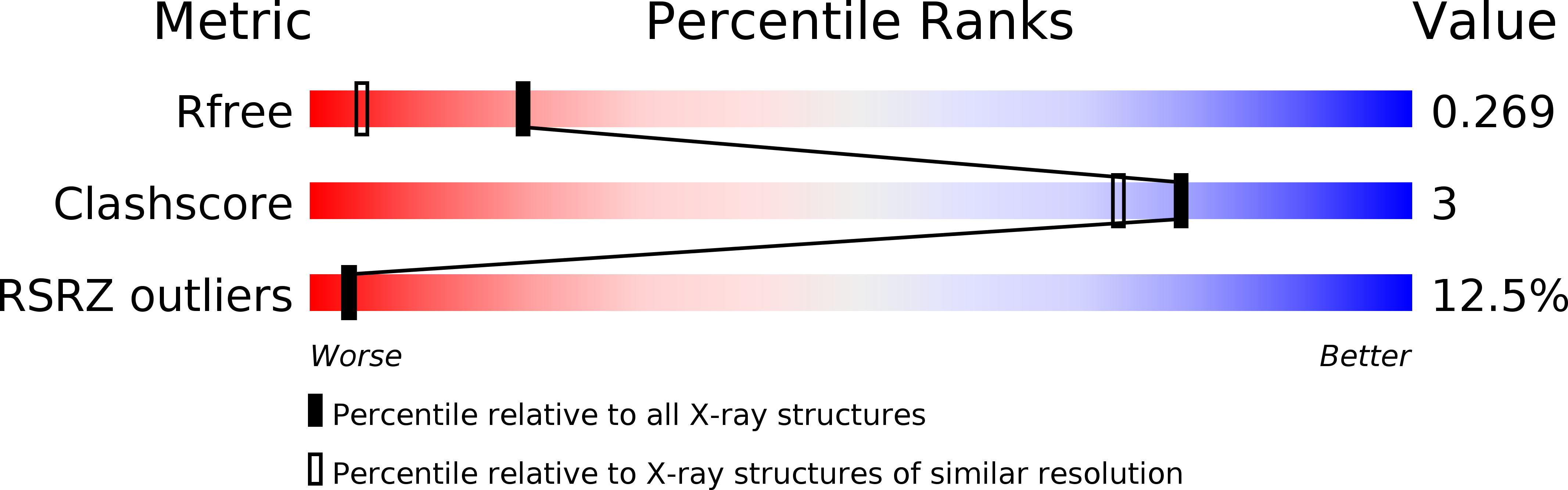
R-Value Free:
0.26
R-Value Work:
0.22
R-Value Observed:
0.22
Space Group:
P 41 2 2