3C0V image
Deposition Date 2008-01-21
Release Date 2008-05-20
Last Version Date 2024-03-13
Entry Detail
PDB ID:
3C0V
Keywords:
Title:
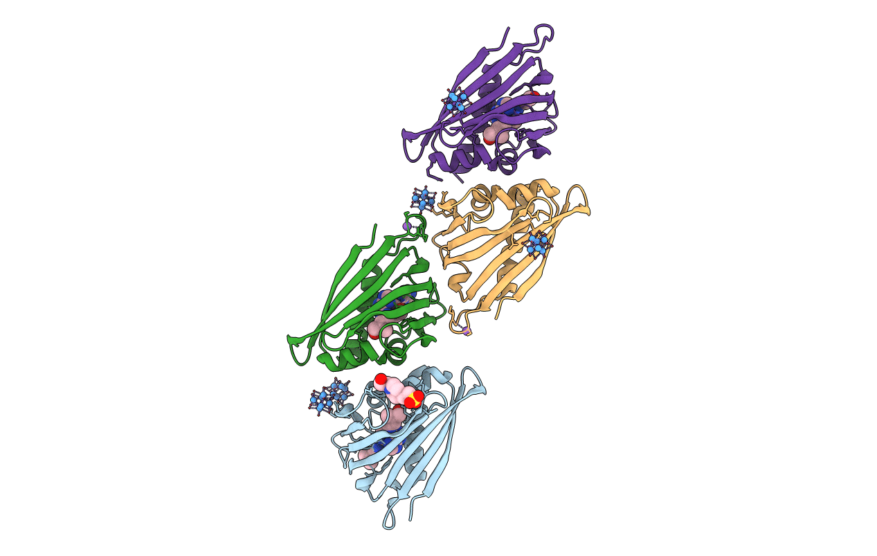
Crystal structure of cytokinin-specific binding protein in complex with cytokinin and Ta6Br12
Biological Source:
Source Organism:
Vigna radiata (Taxon ID: )
Host Organism:
Method Details:
Experimental Method:
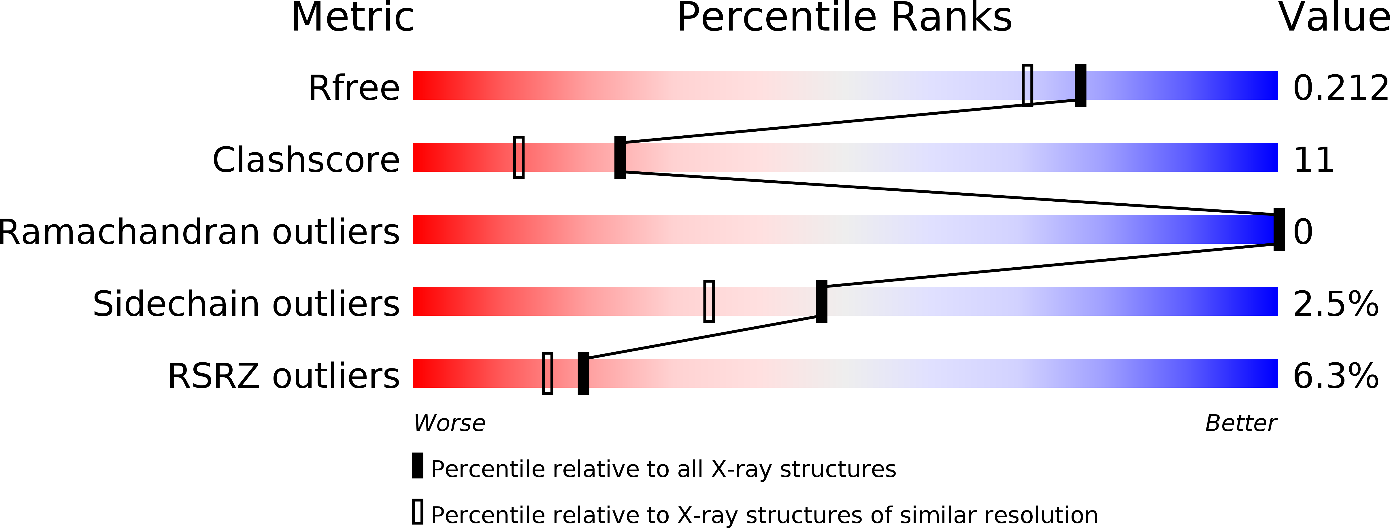
Resolution:
1.80 Å
R-Value Free:
0.20
R-Value Work:
0.15
R-Value Observed:
0.15
Space Group:
P 64
Macromolecular Entities
Polymer Type:polypeptide(L)
Molecule:Cytokinin-specific binding protein
Chain IDs:A, B, C, D
Chain Length:155
Number of Molecules:4
Biological Source:Vigna radiata
Primary Citation
MAD phasing using the (Ta(6)Br(12))(2+) cluster: a retrospective study
Acta Crystallogr.,Sect.D 64 595 606 (2008)
PMID: 18453695 DOI: 10.1107/S0907444908007853

Abstact

The crystal structure of cytokinin-specific binding protein (CSBP) containing four independent molecules with 4 x 155 = 620 residues in the asymmetric unit of the P6(4) unit cell has been solved by three-wavelength MAD using 1.8 angstroms resolution data recorded from a crystal derivatized with the dodecabromohexatantalum cation (Ta6Br12)2+. The diffraction data contained a very strong anomalous signal (allowing successful phasing even using peak SAD data alone) despite the fact that the five (Ta6Br12)2+ clusters found in the asymmetric unit have low occupancy (about 0.3). The derivative structure has been successfully refined to R = 0.158, providing interesting details on the geometry of the (Ta6Br12)2+ cluster, its interactions with the protein and on the backsoaking of a cytokinin ligand that was originally part of a CSBP-cytokinin complex in the native crystals used for (Ta6Br12)2+ derivatization. A simulation analysis of the phasing power of the (Ta6Br12)2+ ions at artificially imposed resolution limits shows that it is not possible to resolve the individual Ta atoms if the dmin limit of the data is higher than 2.9 angstroms. Additionally, for successful Ta identification the (Ta6Br12)2+ complex should be specifically bound and ordered. Good binding at the protein surface is facilitated by the presence of acidic groups, indicating higher pH buffer conditions to be preferable. In addition, the water channels in the crystal should be sufficiently wide (at least 11 angstroms) to allow free diffusion of the (Ta6Br12)2+ ions on soaking. A retrospective look at the initial molecular-replacement calculations provides interesting insights into how the peculiar packing mode and strong bias of the molecular-replacement-phased electron-density maps had hindered successful solution of the structure by this method.

Legend

Protein

Chemical

Disease

Primary Citation of related structures