Deposition Date
2008-01-18
Release Date
2008-04-15
Last Version Date
2024-11-13
Entry Detail
PDB ID:
3C08
Keywords:
Title:
Crystal structure the Fab fragment of matuzumab/EMD72000 (Fab72000)
Biological Source:
Source Organism(s):
Mus musculus (Taxon ID: 10090)
Expression System(s):
Method Details:
Experimental Method:
Resolution:
2.15 Å
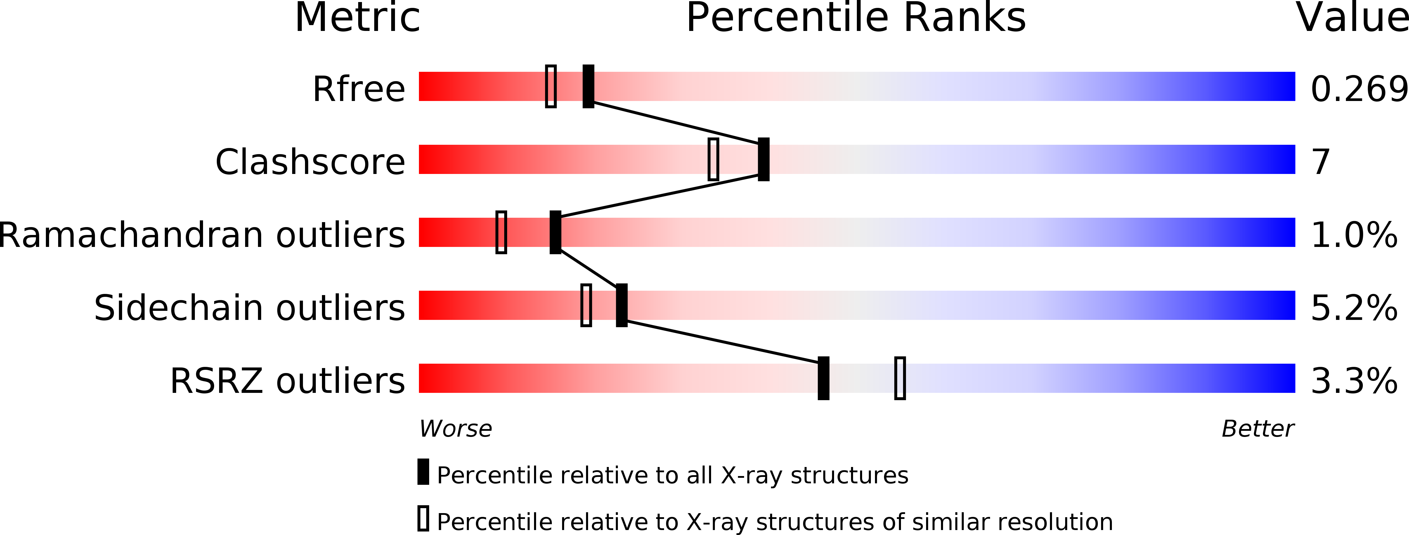
R-Value Free:
0.26
R-Value Work:
0.22
R-Value Observed:
0.22
Space Group:
P 21 21 21