3BXI image
Deposition Date 2008-01-14
Release Date 2008-03-25
Last Version Date 2024-10-16
Entry Detail
PDB ID:
3BXI
Keywords:
Title:
Structure of the complex of bovine lactoperoxidase with its catalyzed product hypothiocyanate ion at 2.3A resolution
Biological Source:
Source Organism:
Bos taurus (Taxon ID: 9913)
Method Details:
Experimental Method:
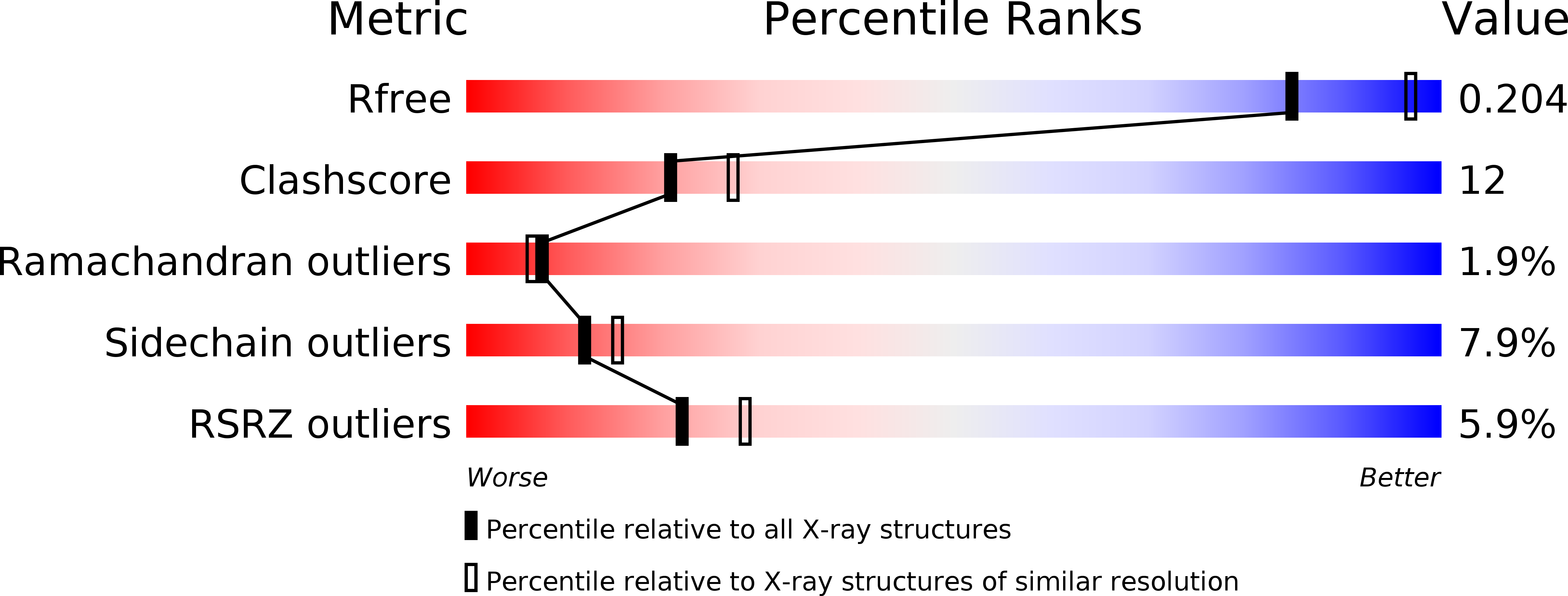
Resolution:
2.30 Å
R-Value Free:
0.21
R-Value Work:
0.16
R-Value Observed:
0.17
Space Group:
P 1 21 1
Macromolecular Entities
Structures with similar UniProt ID
Protein Blast
Polymer Type:polypeptide(L)
Molecule:Lactoperoxidase
Gene (Uniprot):LPO
Chain IDs:A
Chain Length:595
Number of Molecules:1
Biological Source:Bos taurus
Modified Residue
Compound ID Chain ID Parent Comp ID Details 2D Image
ASN A ASN GLYCOSYLATION SITE
SEP A SER PHOSPHOSERINE
Primary Citation
Inhibition of lactoperoxidase by its own catalytic product: crystal structure of the hypothiocyanate-inhibited bovine lactoperoxidase at 2.3-A resolution.
Biophys.J. 96 646 654 (2009)
PMID: 19167310 DOI: 10.1016/j.bpj.2008.09.019

Abstact

To the best of our knowledge, this is the first report on the structure of product-inhibited mammalian peroxidase. Lactoperoxidase is a heme containing an enzyme that catalyzes the inactivation of a wide range of microorganisms. In the presence of hydrogen peroxide, it preferentially converts thiocyanate ion into a toxic hypothiocyanate ion. Samples of bovine lactoperoxidase containing thiocyanate (SCN(-)) and hypothiocyanate (OSCN(-)) ions were purified and crystallized. The structure was determined at 2.3-A resolution and refined to R(cryst) and R(free) factors of 0.184 and 0.221, respectively. The determination of structure revealed the presence of an OSCN(-) ion at the distal heme cavity. The presence of OSCN(-) ions in crystal samples was also confirmed by chemical and spectroscopic analysis. The OSCN(-) ion interacts with the heme iron, Gln-105 N(epsilon1), His-109 N(epsilon2), and a water molecule W96. The sulfur atom of the OSCN(-) ion forms a hypervalent bond with a nitrogen atom of the pyrrole ring D of the heme moiety at an S-N distance of 2.8 A. The heme group is covalently bound to the protein through two ester linkages involving carboxylic groups of Glu-258 and Asp-108 and the modified methyl groups of pyrrole rings A and C, respectively. The heme moiety is significantly distorted from planarity, whereas pyrrole rings A, B, C, and D are essentially planar. The iron atom is displaced by approximately 0.2 A from the plane of the heme group toward the proximal site. The substrate channel resembles a long tunnel whose inner walls contain predominantly aromatic residues such as Phe-113, Phe-239, Phe-254, Phe-380, Phe-381, Phe-422, and Pro-424. A phosphorylated Ser-198 was evident at the surface, in the proximity of the calcium-binding channel.

Legend

Protein

Chemical

Disease

Primary Citation of related structures