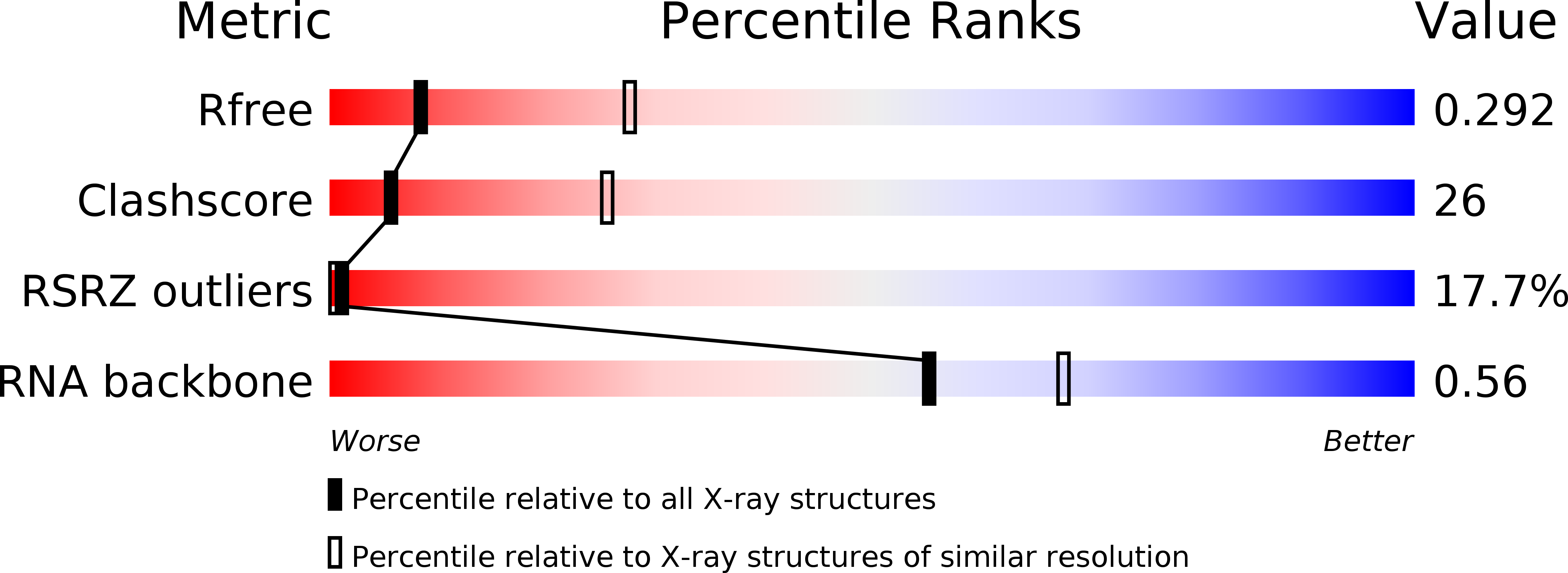
3BWP image
Deposition Date 2008-01-10
Release Date 2008-04-15
Last Version Date 2024-02-21
Entry Detail
PDB ID:
3BWP
Keywords:
Title:
Crystal structure of a self-spliced group II intron
Biological Source:
Source Organism:
(Taxon ID: )
Method Details:
Experimental Method:
Resolution:
3.10 Å
R-Value Free:
0.31
R-Value Work:
0.27
Space Group:
P 21 21 21
Macromolecular Entities
Polymer Type:polyribonucleotide
Molecule:Group IIC intron
Chain IDs:A
Chain Length:412
Number of Molecules:1
Biological Source:
Primary Citation
Crystal structure of a self-spliced group II intron
Science 320 77 82 (2008)
PMID: 18388288 DOI: 10.1126/science.1153803

Abstact

Group II introns are self-splicing ribozymes that catalyze their own excision from precursor transcripts and insertion into new genetic locations. Here we report the crystal structure of an intact, self-spliced group II intron from Oceanobacillus iheyensis at 3.1 angstrom resolution. An extensive network of tertiary interactions facilitates the ordered packing of intron subdomains around a ribozyme core that includes catalytic domain V. The bulge of domain V adopts an unusual helical structure that is located adjacent to a major groove triple helix (catalytic triplex). The bulge and catalytic triplex jointly coordinate two divalent metal ions in a configuration that is consistent with a two-metal ion mechanism for catalysis. Structural and functional analogies support the hypothesis that group II introns and the spliceosome share a common ancestor.

Legend

Protein

Chemical

Disease

Primary Citation of related structures