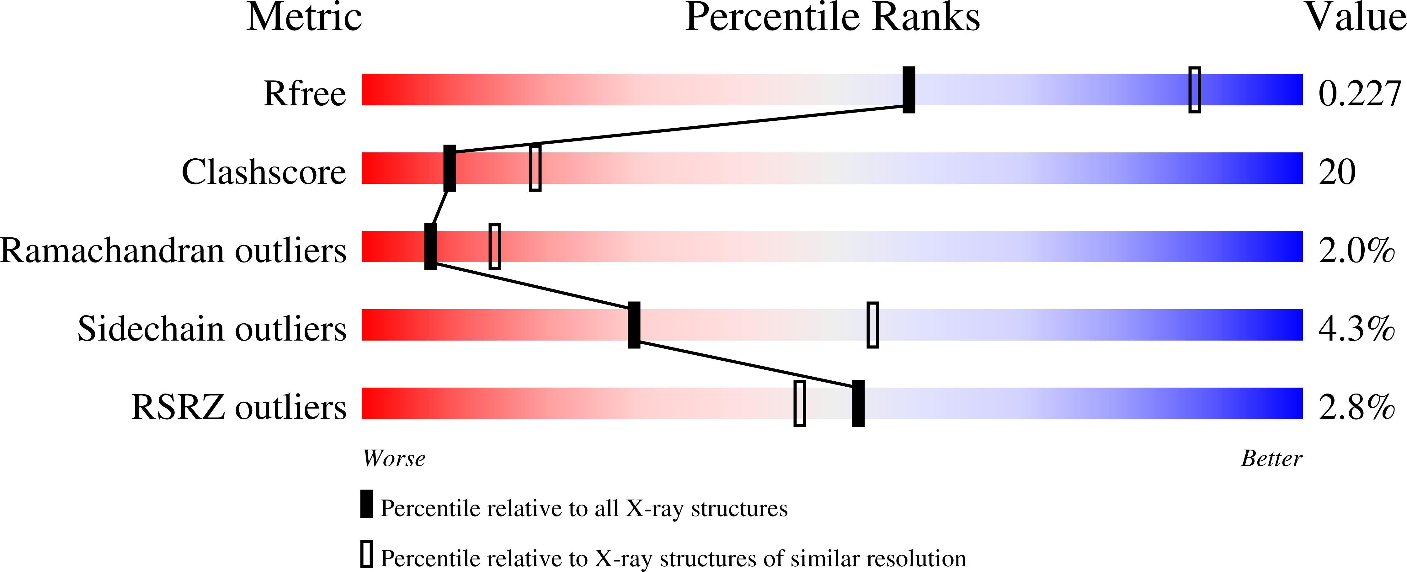
3BVH image
Deposition Date 2008-01-07
Release Date 2008-09-02
Last Version Date 2024-10-30
Entry Detail
PDB ID:
3BVH
Keywords:
Title:
Crystal Structure of Recombinant gammaD364A Fibrinogen Fragment D with the Peptide Ligand Gly-Pro-Arg-Pro-Amide
Biological Source:
Source Organism:
Host Organism:
Method Details:
Experimental Method:
Resolution:
2.60 Å
R-Value Free:
0.26
R-Value Work:
0.22
R-Value Observed:
0.22
Space Group:
P 21 21 21
Macromolecular Entities
Structures with similar UniProt ID
Protein Blast
Polymer Type:polypeptide(L)
Molecule:Fibrinogen alpha chain
Gene (Uniprot):FGA
Chain IDs:A, D
Chain Length:62
Number of Molecules:2
Biological Source:Homo sapiens
Structures with similar UniProt ID
Protein Blast
Polymer Type:polypeptide(L)
Molecule:Fibrinogen beta chain
Gene (Uniprot):FGB
Chain IDs:B, E
Chain Length:298
Number of Molecules:2
Biological Source:Homo sapiens
Structures with similar UniProt ID
Protein Blast
Polymer Type:polypeptide(L)
Molecule:Fibrinogen gamma chain
Gene (Uniprot):FGG
Mutagens:D390A
Chain IDs:C, F
Chain Length:293
Number of Molecules:2
Biological Source:Homo sapiens
Polymer Type:polypeptide(L)
Molecule:4-mer peptide GPRP
Chain IDs:G, H (auth: I), I (auth: H), J
Chain Length:4
Number of Molecules:4
Biological Source:synthetic construct
Modified Residue
Compound ID Chain ID Parent Comp ID Details 2D Image
ASN B ASN GLYCOSYLATION SITE
Ligand Molecules
Primary Citation
Polymerization-defective fibrinogen variant gammaD364A binds knob "A" peptide mimic.
Biochemistry 47 8607 8613 (2008)
PMID: 18642883 DOI: 10.1021/bi8000769

Abstact

Fibrin polymerization is supported in part by interactions called "A:a". Crystallographic studies revealed gamma364Asp is part of hole "a" that interacts with knob "A" peptide mimic, GPRP. Biochemical studies have shown gamma364Asp is critical to polymerization, as polymerization of variants gammaD364A, gammaD364H, and gammaD364V is exceptionally impaired. To understand the molecular basis for the aberrant function, we solved the crystal structure of fragment D from gammaD364A. Surprisingly, the structure (rfD-gammaD364A+GP) showed near normal "A:a" interactions with GPRP bound to hole "a" and no change in the overall structure of gammaD364A. Of note, inspection of the structure showed negative electrostatic potential inside hole "a" was diminished by this substitution. We examined GPRP binding to the gamma364Asp variants in solution by plasmin protection assay. We found no protection of either gammaD364H or gammaD364V but partial protection of gammaD364A, indicating the peptide does not bind to either gammaD364H or gammaD364V and binds more weakly than normal to gammaD364A. We also examined protection by calcium and found all variants were indistinguishable from normal, suggesting the global structures of the variants are not markedly different from normal. Our data imply that gamma364Asp per se is not required for knob "A" binding to hole "a"; rather, this residue's negative charge has a critical role in the electrostatic interactions that facilitate the important first step in fibrin polymerization.

Legend

Protein

Chemical

Disease

Primary Citation of related structures