Deposition Date
2008-01-05
Release Date
2008-04-01
Last Version Date
2023-08-30
Entry Detail
PDB ID:
3BVA
Keywords:
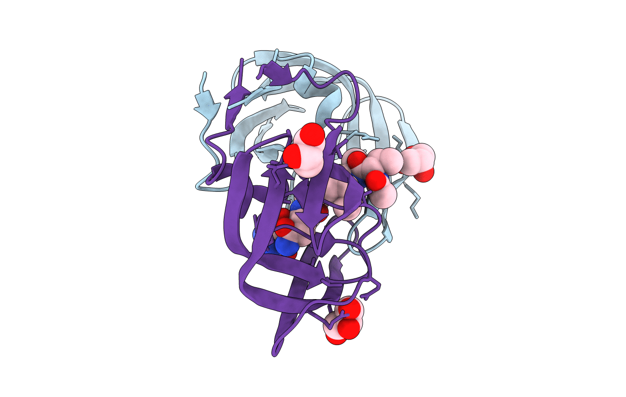
Title:
Cystal structure of HIV-1 Active Site Mutant D25N and p2-NC analog inhibitor
Biological Source:
Source Organism(s):
Human immunodeficiency virus 1 (Taxon ID: 11676)
Expression System(s):
Method Details:
Experimental Method:
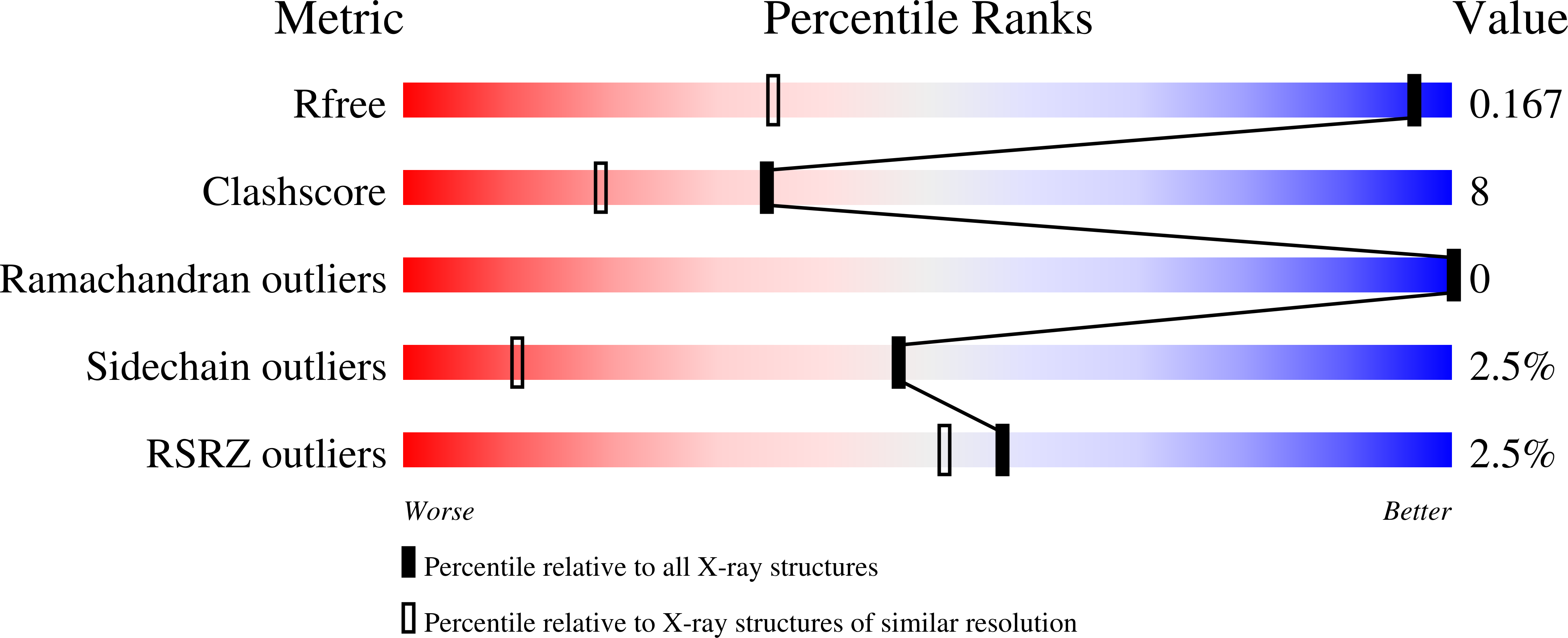
Resolution:
1.05 Å
R-Value Free:
0.18
R-Value Work:
0.15
R-Value Observed:
0.15
Space Group:
P 21 21 2