3BV4 image
Deposition Date 2008-01-04
Release Date 2008-06-24
Last Version Date 2023-08-30
Entry Detail
PDB ID:
3BV4
Keywords:
Title:
Crystal structure of a rabbit muscle fructose-1,6-bisphosphate aldolase A dimer variant
Biological Source:
Source Organism:
Host Organism:
Method Details:
Experimental Method:
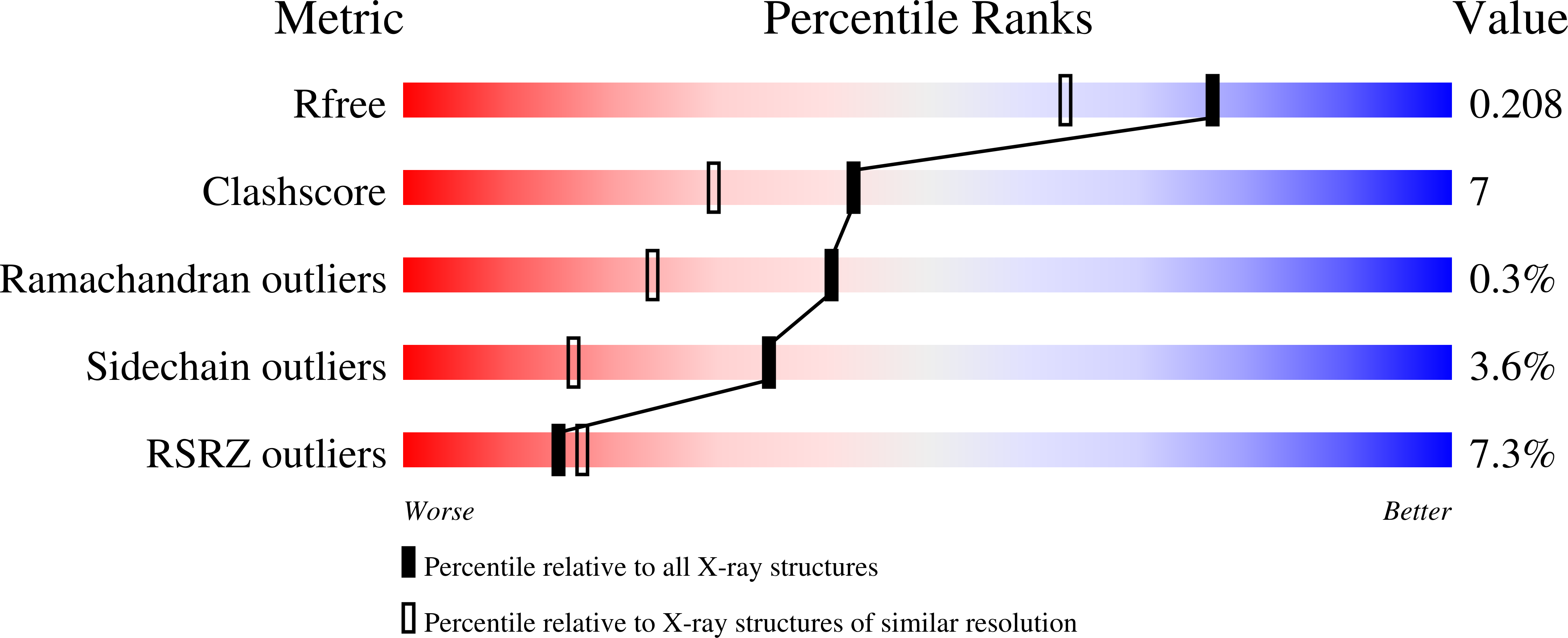
Resolution:
1.70 Å
R-Value Free:
0.21
R-Value Work:
0.19
R-Value Observed:
0.19
Space Group:
P 21 21 2
Macromolecular Entities
Polymer Type:polypeptide(L)
Molecule:Fructose-bisphosphate aldolase A
Gene (Uniprot):ALDOA
Mutations:D128V
Chain IDs:A
Chain Length:341
Number of Molecules:1
Biological Source:Oryctolagus cuniculus
Primary Citation
Structure of a rabbit muscle fructose-1,6-bisphosphate aldolase A dimer variant.
Acta Crystallogr.,Sect.D 64 543 550 (2008)
PMID: 18453690 DOI: 10.1107/S0907444908004976

Abstact

Fructose-1,6-bisphosphate aldolase (aldolase) is an essential enzyme in glycolysis and gluconeogenesis. In addition to this primary function, aldolase is also known to bind to a variety of other proteins, a property that may allow it to perform 'moonlighting' roles in the cell. Although monomeric and dimeric aldolases possess full catalytic activity, the enzyme occurs as an unusually stable tetramer, suggesting a possible link between the oligomeric state and these noncatalytic cellular roles. Here, the first high-resolution X-ray crystal structure of rabbit muscle D128V aldolase, a dimeric form of aldolase mimicking the clinically important D128G mutation in humans associated with hemolytic anemia, is presented. The structure of the dimer was determined to 1.7 angstroms resolution with the product DHAP bound in the active site. The turnover of substrate to produce the product ligand demonstrates the retention of catalytic activity by the dimeric aldolase. The D128V mutation causes aldolase to lose intermolecular contacts with the neighboring subunit at one of the two interfaces of the tetramer. The tertiary structure of the dimer does not significantly differ from the structure of half of the tetramer. Analytical ultracentrifugation confirms the occurrence of the enzyme as a dimer in solution. The highly stable structure of aldolase with an independent active site is consistent with a model in which aldolase has evolved as a multimeric scaffold to perform other noncatalytic functions.

Legend

Protein

Chemical

Disease

Primary Citation of related structures