3BUZ image
Deposition Date 2008-01-04
Release Date 2008-05-13
Last Version Date 2023-11-01
Entry Detail
PDB ID:
3BUZ
Title:
Crystal structure of ia-bTAD-actin complex
Biological Source:
Source Organism:
Host Organism:
Method Details:
Experimental Method:
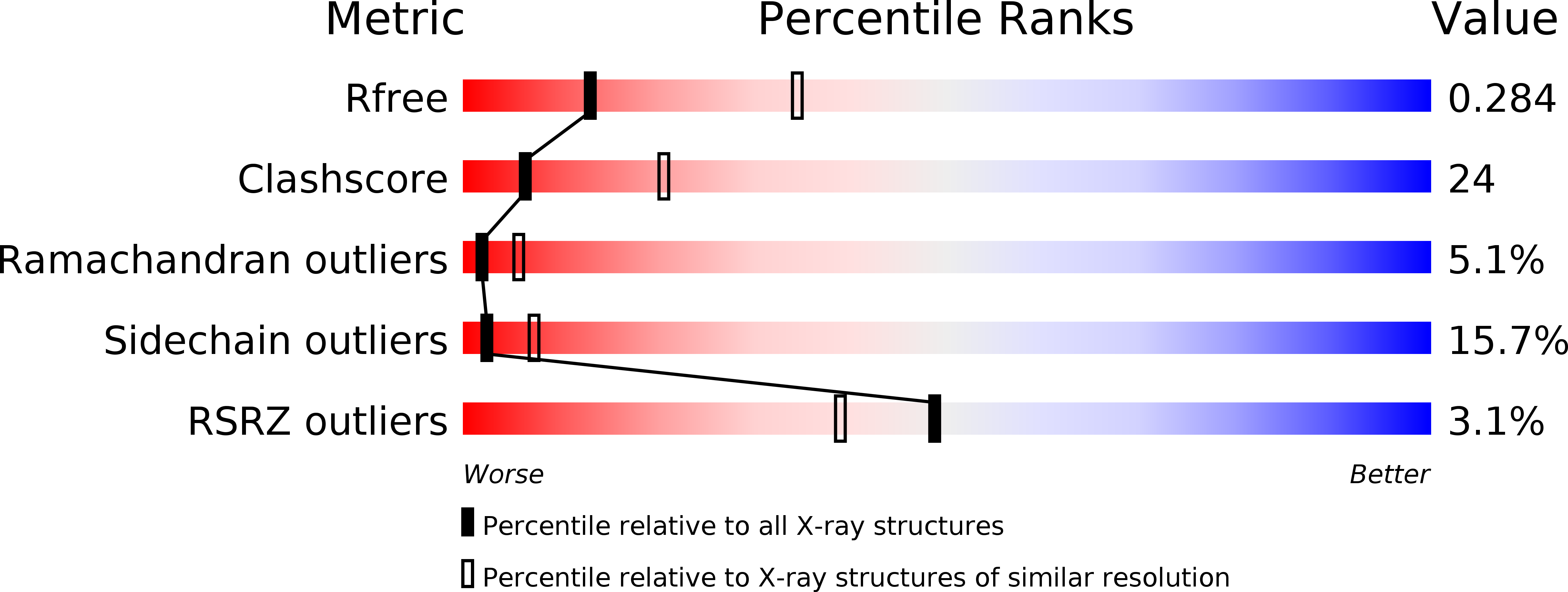
Resolution:
2.81 Å
R-Value Free:
0.29
R-Value Work:
0.22
R-Value Observed:
0.22
Space Group:
P 21 21 21
Macromolecular Entities
Polymer Type:polypeptide(L)
Molecule:Iota toxin component Ia
Chain IDs:A
Chain Length:413
Number of Molecules:1
Biological Source:Clostridium perfringens
Polymer Type:polypeptide(L)
Molecule:Actin, alpha skeletal muscle
Gene (Uniprot):ACTA1
Chain IDs:B
Chain Length:375
Number of Molecules:1
Biological Source:Oryctolagus cuniculus
Primary Citation
Structural basis of actin recognition and arginine ADP-ribosylation by Clostridium perfringens iota-toxin
Proc.Natl.Acad.Sci.Usa 105 7399 7404 (2008)
PMID: 18490658 DOI: 10.1073/pnas.0801215105

Abstact

The ADP-ribosylating toxins (ADPRTs) produced by pathogenic bacteria modify intracellular protein and affect eukaryotic cell function. Actin-specific ADPRTs (including Clostridium perfringens iota-toxin and Clostridium botulinum C2 toxin) ADP-ribosylate G-actin at Arg-177, leading to disorganization of the cytoskeleton and cell death. Although the structures of many actin-specific ADPRTs are available, the mechanisms underlying actin recognition and selective ADP-ribosylation of Arg-177 remain unknown. Here we report the crystal structure of actin-Ia in complex with the nonhydrolyzable NAD analog betaTAD at 2.8 A resolution. The structure indicates that Ia recognizes actin via five loops around NAD: loop I (Tyr-60-Tyr-62 in the N domain), loop II (active-site loop), loop III, loop IV (PN loop), and loop V (ADP-ribosylating turn-turn loop). We used site-directed mutagenesis to confirm that loop I on the N domain and loop II are essential for the ADP-ribosyltransferase activity. Furthermore, we revealed that Glu-378 on the EXE loop is in close proximity to Arg-177 in actin, and we proposed that the ADP-ribosylation of Arg-177 proceeds by an SN1 reaction via first an oxocarbenium ion intermediate and second a cationic intermediate by alleviating the strained conformation of the first oxocarbenium ion. Our results suggest a common reaction mechanism for ADPRTs. Moreover, the structure might be of use in rational drug design to block toxin-substrate recognition.

Legend

Protein

Chemical

Disease

Primary Citation of related structures