
Deposition Date
2008-01-03
Release Date
2008-04-01
Last Version Date
2023-08-30
Entry Detail
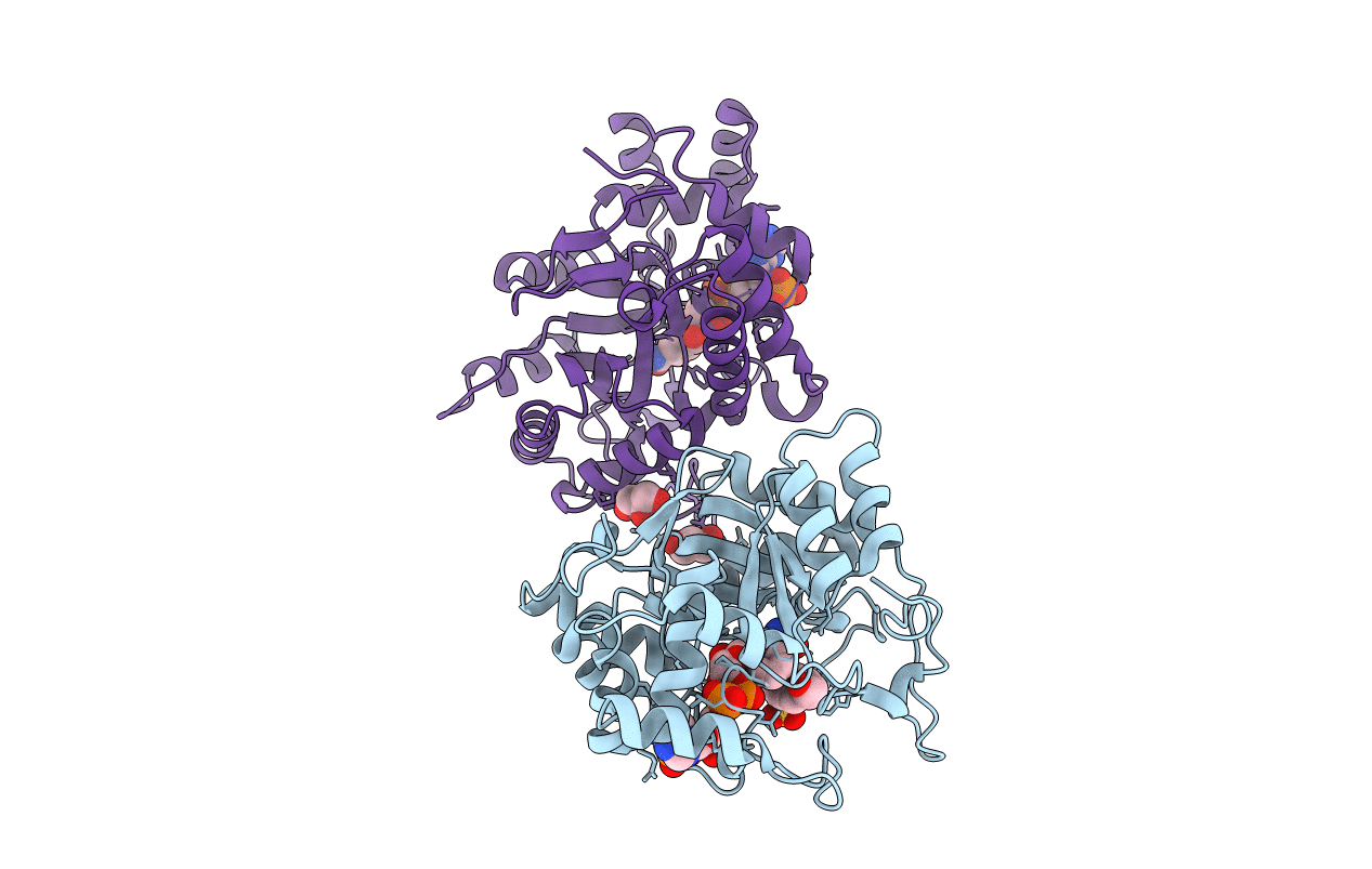
PDB ID:
3BUV
Keywords:
Title:
Crystal structure of human Delta(4)-3-ketosteroid 5-beta-reductase in complex with NADP and HEPES. Resolution: 1.35 A.
Biological Source:
Source Organism(s):
Homo sapiens (Taxon ID: 9606)
Expression System(s):
Method Details:
Experimental Method:
Resolution:
1.35 Å
R-Value Free:
0.23
R-Value Work:
0.21
R-Value Observed:
0.21
Space Group:
P 21 21 21
