3BUJ image
Deposition Date 2008-01-02
Release Date 2008-04-29
Last Version Date 2023-08-30
Entry Detail
PDB ID:
3BUJ
Title:
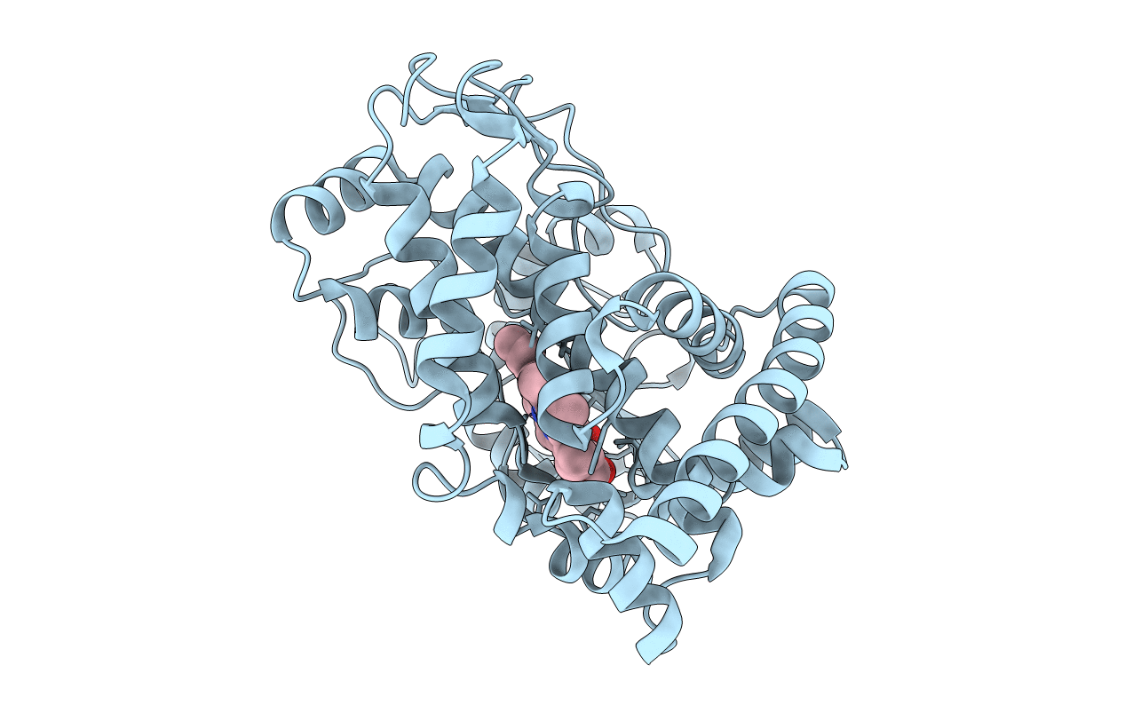
Crystal Structure of CalO2
Biological Source:
Source Organism:
Host Organism:
Method Details:
Experimental Method:
Resolution:
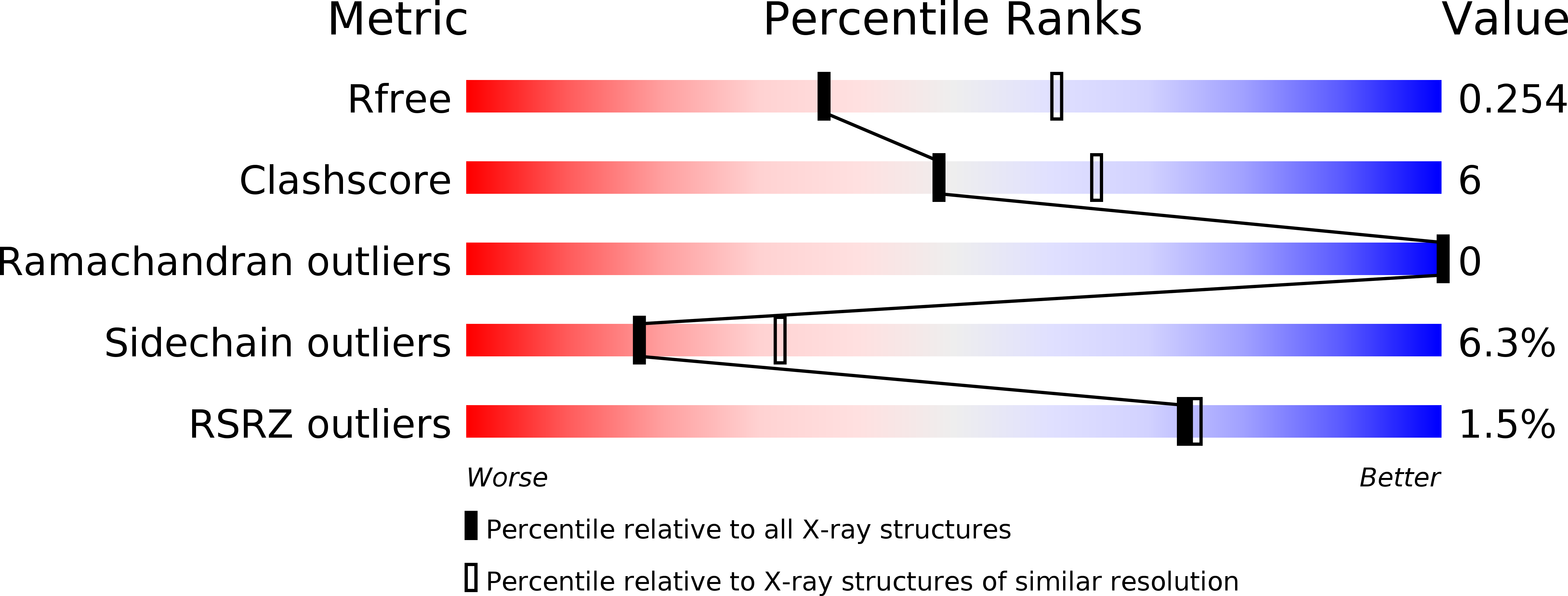
2.47 Å
R-Value Free:
0.25
R-Value Work:
0.19
R-Value Observed:
0.20
Space Group:
P 41 21 2
Macromolecular Entities
Structures with similar UniProt ID
Protein Blast
Polymer Type:polypeptide(L)
Molecule:CalO2
Gene (Uniprot):calO2
Chain IDs:A
Chain Length:397
Number of Molecules:1
Biological Source:Micromonospora echinospora
Ligand Molecules
Primary Citation
Structural characterization of CalO2: a putative orsellinic acid P450 oxidase in the calicheamicin biosynthetic pathway.
Proteins 74 50 60 (2009)
PMID: 18561189 DOI: 10.1002/prot.22131

Abstact

Although bacterial iterative Type I polyketide synthases are now known to participate in the biosynthesis of a small set of diverse natural products, the subsequent downstream modification of the resulting polyketide products remains poorly understood. Toward this goal, we report the X-ray structure determination at 2.5 A resolution and preliminary characterization of the putative orsellenic acid P450 oxidase (CalO2) involved in calicheamicin biosynthesis. These studies represent the first crystal structure for a P450 involved in modifying a bacterial iterative Type I polyketide product and suggest the CalO2-catalyzed step may occur after CalO3-catalyzed iodination and may also require a coenzyme A- (CoA) or acyl carrier protein- (ACP) bound substrate. Docking studies also reveal a putative docking site within CalO2 for the CLM orsellinic acid synthase (CalO5) ACP domain which involves a well-ordered helix along the CalO2 active site cavity that is unique compared with other P450 structures.

Legend

Protein

Chemical

Disease

Primary Citation of related structures