3BT7 image
Deposition Date 2007-12-27
Release Date 2008-04-29
Last Version Date 2024-12-25
Entry Detail
PDB ID:
3BT7
Keywords:
Title:
Structure of E. coli 5-Methyluridine Methyltransferase TrmA in complex with 19 nucleotide T-arm analogue
Biological Source:
Source Organism:
Method Details:
Experimental Method:
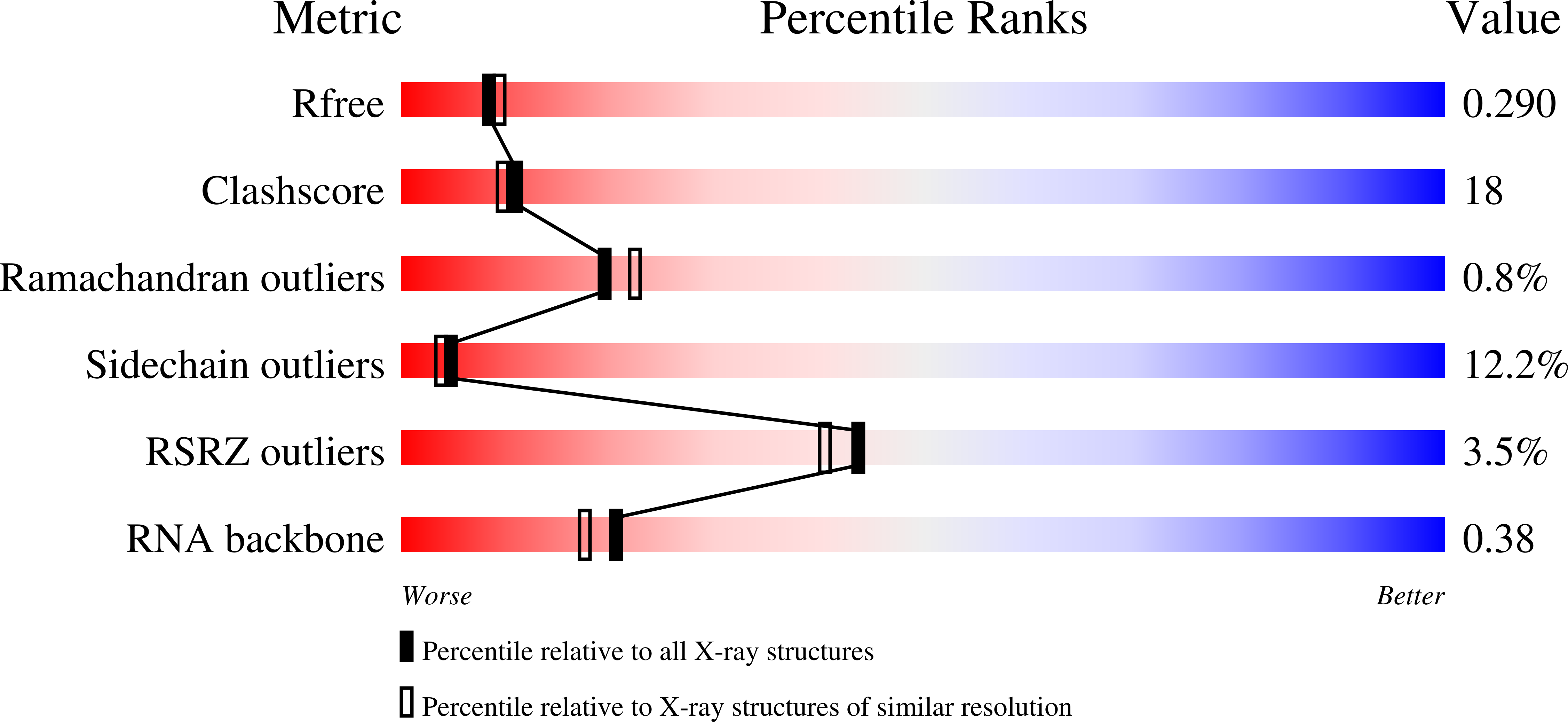
Resolution:
2.43 Å
R-Value Free:
0.28
R-Value Work:
0.22
R-Value Observed:
0.22
Space Group:
C 1 2 1
Macromolecular Entities
Structures with similar UniProt ID
Protein Blast
Polymer Type:polypeptide(L)
Molecule:tRNA (uracil-5-)-methyltransferase
Gene (Uniprot):trmA
Mutagens:E358Q
Chain IDs:A, B
Chain Length:369
Number of Molecules:2
Biological Source:Escherichia coli
Polymer Type:polyribonucleotide
Molecule:RNA (5'-D(P*GP*CP*UP*GP*UP*GP*(5MU)P*UP*CP*GP*AP*UP*CP*CP*AP*CP*AP*GP*C)-3')
Chain IDs:C, D
Chain Length:19
Number of Molecules:2
Biological Source:synthetic construct
Modified Residue
Compound ID Chain ID Parent Comp ID Details 2D Image
5MU C U 5-METHYLURIDINE 5'-MONOPHOSPHATE
Primary Citation
Structure of a TrmA-RNA complex: A consensus RNA fold contributes to substrate selectivity and catalysis in m5U methyltransferases.
Proc.Natl.Acad.Sci.Usa 105 6876 6881 (2008)
PMID: 18451029 DOI: 10.1073/pnas.0802247105

Abstact

TrmA catalyzes S-adenosylmethionine (AdoMet)-dependent methylation of U54 in most tRNAs. We solved the structure of the Escherichia coli 5-methyluridine (m(5)U) 54 tRNA methyltransferase (MTase) TrmA in a covalent complex with a 19-nt T arm analog to 2.4-A resolution. Mutation of the TrmA catalytic base Glu-358 to Gln arrested catalysis and allowed isolation of the covalent TrmA-RNA complex for crystallization. The protein-RNA interface includes 6 nt of the T loop and two proximal base pairs of the stem. U54 is flipped out of the loop into the active site. A58 occupies the space of the everted U54 and is part of a collinear base stack G53-A58-G57-C56-U55. The RNA fold is different from T loop conformations in unbound tRNA or T arm analogs, but nearly identical to the fold of the RNA loop bound at the active site of the m(5)U MTase RumA. In both enzymes, this consensus fold presents the target U and the following two bases to a conserved binding groove on the protein. Outside of this fold, the RumA and TrmA substrates have completely different structures and protein interfaces. Loop residues other than the target U54 make more than half of their hydrogen bonds to the protein via sugar-phosphate moieties, accounting, in part, for the broad consensus sequence for TrmA substrates.

Legend

Protein

Chemical

Disease

Primary Citation of related structures
Feedback Form
Name
Email
Institute
Feedback