Deposition Date
2007-12-27
Release Date
2008-03-25
Last Version Date
2024-11-20
Entry Detail
PDB ID:
3BT1
Keywords:
Title:
Structure of urokinase receptor, urokinase and vitronectin complex
Biological Source:
Source Organism(s):
Homo sapiens (Taxon ID: 9606)
Expression System(s):
Method Details:
Experimental Method:
Resolution:
2.80 Å
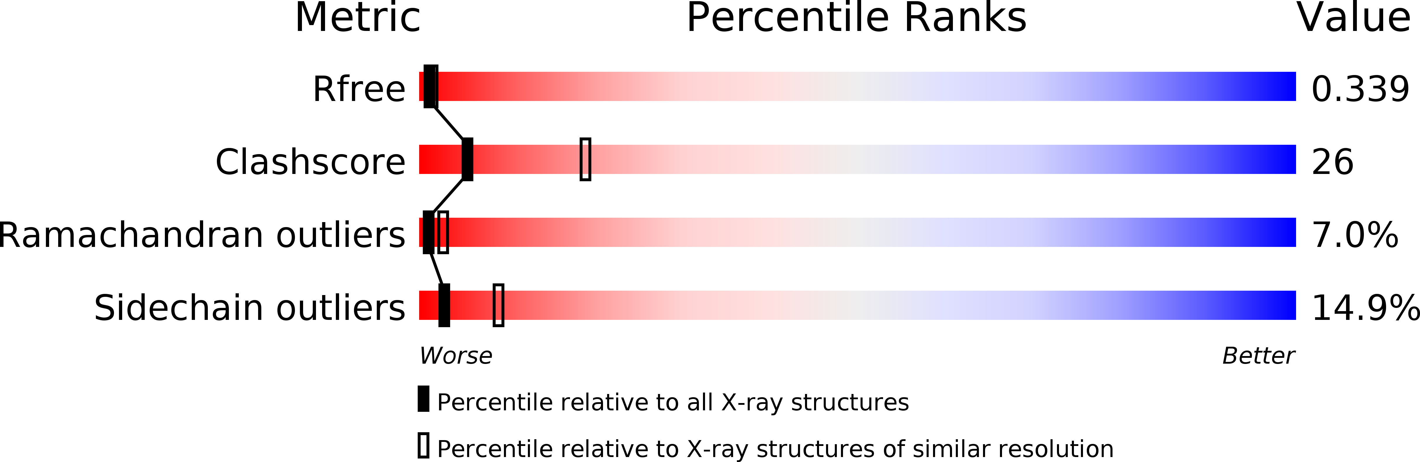
R-Value Free:
0.30
R-Value Work:
0.24
R-Value Observed:
0.24
Space Group:
P 21 21 2