3BSZ image
Deposition Date 2007-12-27
Release Date 2008-11-11
Last Version Date 2024-11-13
Entry Detail
PDB ID:
3BSZ
Title:
Crystal structure of the transthyretin-retinol binding protein-Fab complex
Biological Source:
Source Organism:
Homo sapiens (Taxon ID: 9606)
Mus musculus (Taxon ID: 10090)
Host Organism:
Method Details:
Experimental Method:
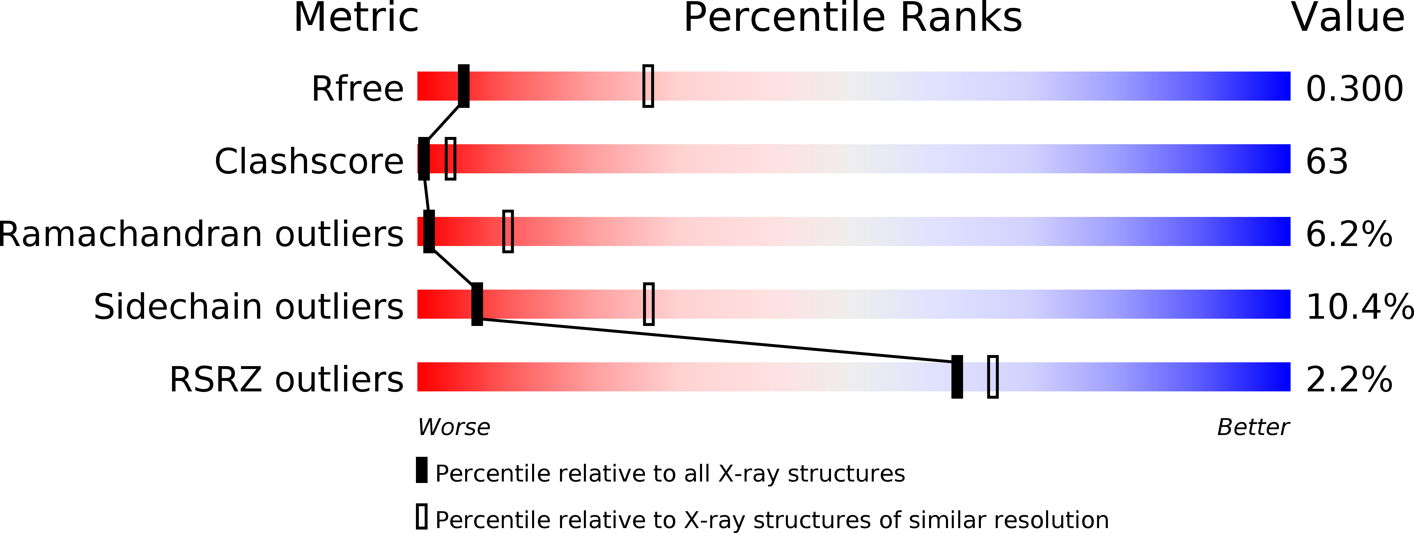
Resolution:
3.38 Å
R-Value Free:
0.31
R-Value Work:
0.23
R-Value Observed:
0.23
Space Group:
C 2 2 2
Macromolecular Entities
Structures with similar UniProt ID
Protein Blast
Polymer Type:polypeptide(L)
Molecule:Transthyretin
Gene (Uniprot):TTR
Chain IDs:A, B, C, D
Chain Length:127
Number of Molecules:4
Biological Source:Homo sapiens
Structures with similar UniProt ID
Protein Blast
Polymer Type:polypeptide(L)
Molecule:Plasma retinol-binding protein
Gene (Uniprot):RBP4
Chain IDs:E, F
Chain Length:176
Number of Molecules:2
Biological Source:Homo sapiens
Polymer Type:polypeptide(L)
Molecule:Fab fragment light chain
Chain IDs:H, J (auth: N)
Chain Length:215
Number of Molecules:2
Biological Source:Mus musculus
Polymer Type:polypeptide(L)
Molecule:Fab fragment heavy chain
Chain IDs:G (auth: L), I (auth: M)
Chain Length:215
Number of Molecules:2
Biological Source:Mus musculus
Ligand Molecules
Primary Citation
Structural and mutational analyses of protein-protein interactions between transthyretin and retinol-binding protein.
Febs J. 275 5841 5854 (2008)
PMID: 19021760 DOI: 10.1111/j.1742-4658.2008.06705.x

Abstact

Transthyretin is a tetrameric binding protein involved in the transport of thyroid hormones and in the cotransport of retinol by forming a complex in plasma with retinol-binding protein. In the present study, we report the crystal structure of a macromolecular complex, in which human transthyretin, human holo-retinol-binding protein and a murine anti-retinol-binding protein Fab are assembled according to a 1 : 2 : 2 stoichiometry. The main interactions, both polar and apolar, between retinol-binding protein and transthyretin involve the retinol hydroxyl group and a limited number of solvent exposed residues. The relevance of transthyretin residues in complex formation with retinol-binding protein has been examined by mutational analysis, and the structural consequences of some transthyretin point mutations affecting protein-protein recognition have been investigated. Despite a few exceptions, in general, the substitution of a hydrophilic for a hydrophobic side chain in contact regions results in a decrease or even a loss of binding affinity, thus revealing the importance of interfacial hydrophobic interactions and a high degree of complementarity between retinol-binding protein and transthyretin. The effect is particularly evident when the mutation affects an interacting residue present in two distinct subunits of transthyretin participating simultaneously in two interactions with a retinol-binding protein molecule. This is the case of the amyloidogenic I84S replacement, which abolishes the interaction with retinol-binding protein and is associated with an altered retinol-binding protein plasma transport in carriers of this mutation. Remarkably, some of the residues in mutated human transthyretin that weaken or abolish the interaction with retinol-binding protein are present in piscine transthyretin, consistent with the lack of interaction between retinol-binding protein and transthyretin in fish.

Legend

Protein

Chemical

Disease

Primary Citation of related structures