3BSW image
Entry Detail
PDB ID:
3BSW
Keywords:
Title:
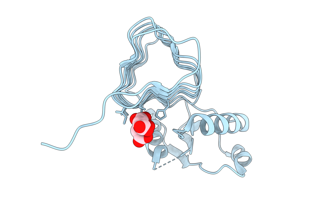
PglD-citrate complex, from Campylobacter jejuni NCTC 11168
Biological Source:
Source Organism:
Host Organism:
PDB Version:
Deposition Date:
2007-12-26
Release Date:
2008-07-29
Method Details:
Experimental Method:
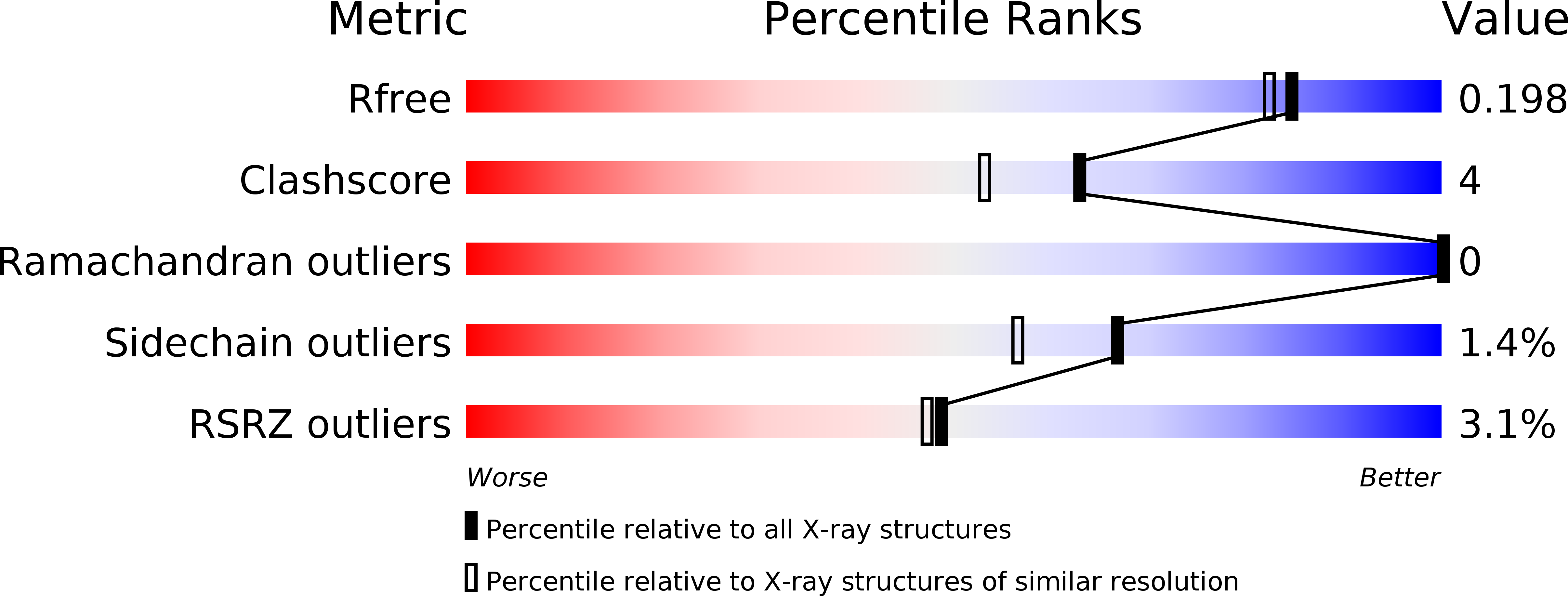
Resolution:
1.77 Å
R-Value Free:
0.19
R-Value Work:
0.18
R-Value Observed:
0.18
Space Group:
P 63
Macromolecular Entities
Polymer Type:polypeptide(L)
Description:Acetyltransferase
Chain IDs:A
Chain Length:198
Number of Molecules:1
Biological Source:Campylobacter jejuni
Ligand Molecules
Primary Citation
Crystal structure and catalytic mechanism of PglD from Campylobacter jejuni.
J.Biol.Chem. 283 27937 27946 (2008)
PMID: 18667421 DOI: 10.1074/jbc.M801207200

Abstact

The carbohydrate 2, 4-diacetamido-2, 4, 6-trideoxy-alpha-D-glucopyranose (BacAc(2)) is found in a variety of eubacterial pathogens. In Campylobacter jejuni, PglD acetylates the C4 amino group on UDP-2-acetamido-4-amino-2, 4, 6-trideoxy-alpha-D-glucopyranose (UDP-4-amino-sugar) to form UDP-BacAc(2). Sequence analysis predicts PglD to be a member of the left-handed beta helix family of enzymes. However, poor sequence homology between PglD and left-handed beta helix enzymes with existing structural data precludes unambiguous identification of the active site. The co-crystal structures of PglD in the presence of citrate, acetyl coenzyme A, or the UDP-4-amino-sugar were solved. The biological assembly is a trimer with one active site formed between two protomers. Residues lining the active site were identified, and results from functional assays on alanine mutants suggest His-125 is critical for catalysis, whereas His-15 and His-134 are involved in substrate binding. These results are discussed in the context of implications for proteins homologous to PglD in other pathogens.

Legend

Protein

Chemical

Disease

Primary Citation of related structures