3BSU image
Entry Detail
PDB ID:
3BSU
Title:
Hybrid-binding domain of human RNase H1 in complex with 12-mer RNA/DNA
Biological Source:
Source Organism:
Host Organism:
PDB Version:
Deposition Date:
2007-12-26
Release Date:
2008-03-25
Method Details:
Experimental Method:
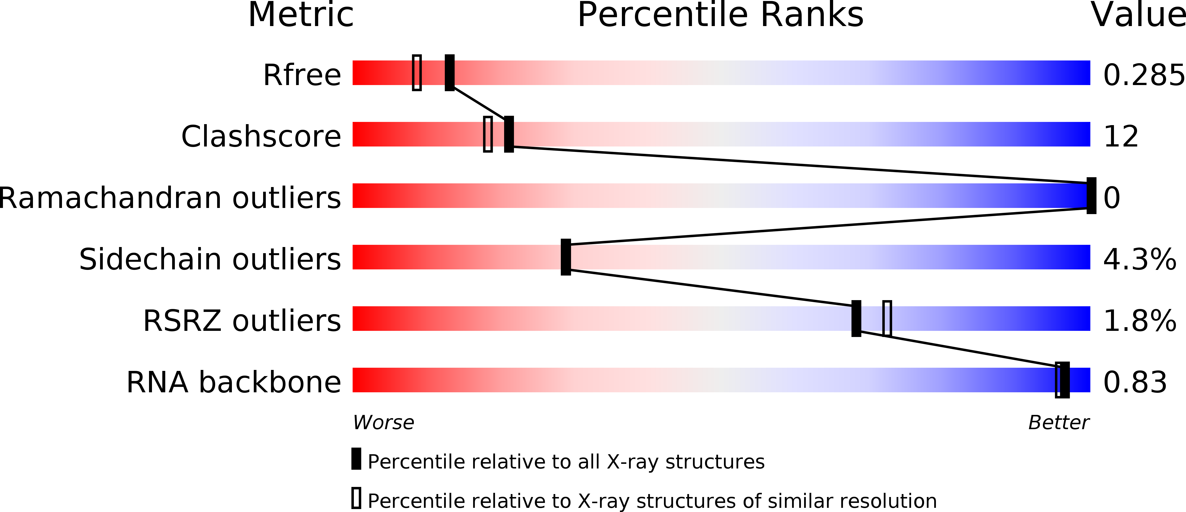
Resolution:
2.10 Å
R-Value Free:
0.29
R-Value Work:
0.22
Space Group:
P 21 21 21
Macromolecular Entities
Polymer Type:polypeptide(L)
Description:Ribonuclease H1
Chain IDs:E (auth: A), F (auth: B), G (auth: C), H (auth: F), I (auth: G), J (auth: H)
Chain Length:53
Number of Molecules:6
Biological Source:Homo sapiens
Modified Residue
Compound ID Chain ID Parent Comp ID Details 2D Image
5IU B DU 5-IODO-2'-DEOXYURIDINE-5'-MONOPHOSPHATE
Ligand Molecules
Primary Citation
Specific recognition of RNA/DNA hybrid and enhancement of human RNase H1 activity by HBD.
Embo J. 27 1172 1181 (2008)
PMID: 18337749 DOI: 10.1038/emboj.2008.44

Abstact

Human RNase H1 contains an N-terminal domain known as dsRHbd for binding both dsRNA and RNA/DNA hybrid. We find that dsRHbd binds preferentially to RNA/DNA hybrids by over 25-fold and rename it as hybrid binding domain (HBD). The crystal structure of HBD complexed with a 12 bp RNA/DNA hybrid reveals that the RNA strand is recognized by a protein loop, which forms hydrogen bonds with the 2'-OH groups. The DNA interface is highly specific and contains polar residues that interact with the phosphate groups and an aromatic patch that appears selective for binding deoxyriboses. HBD is unique relative to non-sequence-specific dsDNA- and dsRNA-binding domains because it does not use positive dipoles of alpha-helices for nucleic acid binding. Characterization of full-length enzymes with defective HBDs indicates that this domain dramatically enhances both the specific activity and processivity of RNase H1. Similar activity enhancement by small substrate-binding domains linked to the catalytic domain likely occurs in other nucleic acid enzymes.

Legend

Protein

Chemical

Disease

Primary Citation of related structures