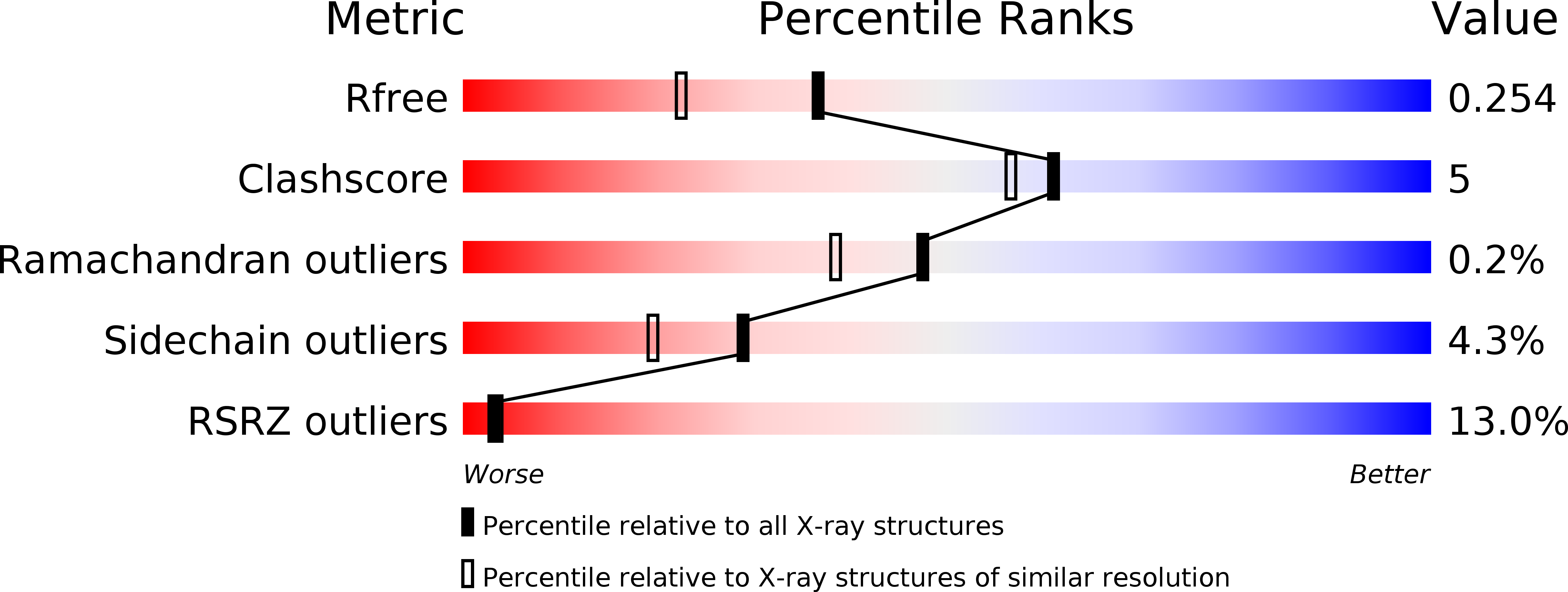
3BRB image
Deposition Date 2007-12-21
Release Date 2008-01-22
Last Version Date 2023-08-30
Entry Detail
PDB ID:
3BRB
Keywords:
Title:
Crystal structure of catalytic domain of the proto-oncogene tyrosine-protein kinase MER in complex with ADP
Biological Source:
Source Organism:
Homo sapiens (Taxon ID: 9606)
Method Details:
Experimental Method:
Resolution:
1.90 Å
R-Value Free:
0.24
R-Value Work:
0.19
R-Value Observed:
0.19
Space Group:
P 1 21 1
Macromolecular Entities
Structures with similar UniProt ID
Protein Blast
Polymer Type:polypeptide(L)
Molecule:Proto-oncogene tyrosine-protein kinase MER
Gene (Uniprot):MERTK
Chain IDs:A, B
Chain Length:313
Number of Molecules:2
Biological Source:Homo sapiens
Primary Citation
Structural insights into the inhibited states of the Mer receptor tyrosine kinase.
J.Struct.Biol. 165 88 96 (2009)
PMID: 19028587 DOI: 10.1016/j.jsb.2008.10.003

Abstact

The mammalian ortholog of the retroviral oncogene v-Eyk, and a receptor tyrosine kinase upstream of antiapoptotic and transforming signals, Mer (MerTK) is a mediator of the phagocytic process, being involved in retinal and immune cell clearance and platelet aggregation. Mer knockout mice are viable and are protected from epinephrine-induced pulmonary thromboembolism and ferric chloride-induced thrombosis. Mer overexpression, on the other hand, is associated with numerous carcinomas. Although Mer adaptor proteins and signaling pathways have been identified, it remains unclear how Mer initiates phagocytosis. When bound to its nucleotide cofactor, the high-resolution structure of Mer shows an autoinhibited alphaC-Glu-out conformation with insertion of an activation loop residue into the active site. Mer complexed with compound-52 (C52: 2-(2-hydroxyethylamino)-6-(3-chloroanilino)-9-isopropylpurine), a ligand identified from a focused library, retains its DFG-Asp-in and alphaC-Glu-out conformation, but acquires other conformational changes. The alphaC helix and DFGL region is closer to the hinge region and the ethanolamine moiety of C52 binds in the groove formed between Leu593 and Val601 of the P-loop, causing a compression of the active site pocket. These conformational states reveal the mechanisms of autoinhibition, the pathophysiological basis of disease-causing mutations, and a platform for the development of chemical probes.

Legend

Protein

Chemical

Disease

Primary Citation of related structures