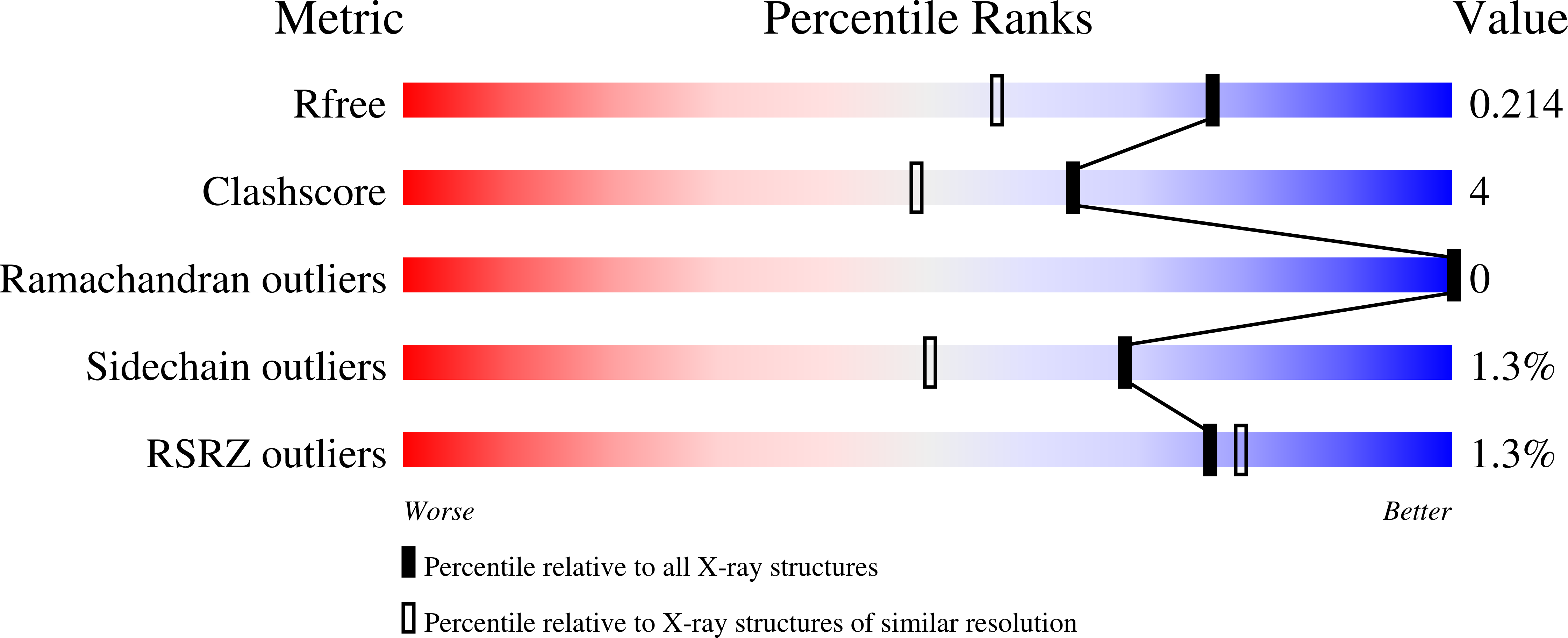
3BPZ image
Deposition Date 2007-12-19
Release Date 2008-03-25
Last Version Date 2023-08-30
Entry Detail
PDB ID:
3BPZ
Title:
HCN2-I 443-460 E502K in the presence of cAMP
Biological Source:
Source Organism:
Mus musculus (Taxon ID: 10090)
Method Details:
Experimental Method:
Resolution:
1.65 Å
R-Value Free:
0.21
R-Value Work:
0.19
R-Value Observed:
0.19
Space Group:
C 2 2 21
Macromolecular Entities
Polymer Type:polypeptide(L)
Molecule:Potassium/sodium hyperpolarization-activated cyclic nucleotide-gated channel 2
Gene (Uniprot):Hcn2
Mutations:E502K
Chain IDs:A, B, C, D
Chain Length:202
Number of Molecules:4
Biological Source:Mus musculus
Ligand Molecules
Primary Citation
C-terminal movement during gating in cyclic nucleotide-modulated channels.
J.Biol.Chem. 283 14728 14738 (2008)
PMID: 18367452 DOI: 10.1074/jbc.M710463200

Abstact

Activation of cyclic nucleotide-modulated channels such as CNG and HCN channels is promoted by ligand-induced conformational changes in their C-terminal regions. The primary intersubunit interface of these C termini includes two salt bridges per subunit, formed between three residues (one positively charged and two negatively charged amino acids) that we term the SB triad. We previously hypothesized that the SB triad is formed in the closed channel and breaks when the channel opens. Here we tested this hypothesis by dynamically manipulating the SB triad in functioning CNGA1 channels. Reversing the charge at positions Arg-431 and Glu-462, two of the SB triad residues, by either mutation or application of charged reagents increased the favorability of channel opening. To determine how a charge reversal mutation in the SB triad structurally affects the channel, we solved the crystal structure of the HCN2 C-terminal region with the equivalent E462R mutation. The backbone structure of this mutant was very similar to that of wild type, but the SB triad was rearranged such that both salt bridges did not always form simultaneously, suggesting a mechanism for the increased ease of opening of the mutant channels. To prevent movement in the SB triad, we tethered two components of the SB triad region together with cysteine-reactive cross-linkers. Preventing normal movement of the SB triad region with short cross-linkers inhibited channel opening, whereas longer cross-linkers did not. These results support our hypothesis that the SB triad forms in the closed channel and indicate that this region expands as the channel opens.

Legend

Protein

Chemical

Disease

Primary Citation of related structures