3BP3 image
Entry Detail
PDB ID:
3BP3
Keywords:
Title:
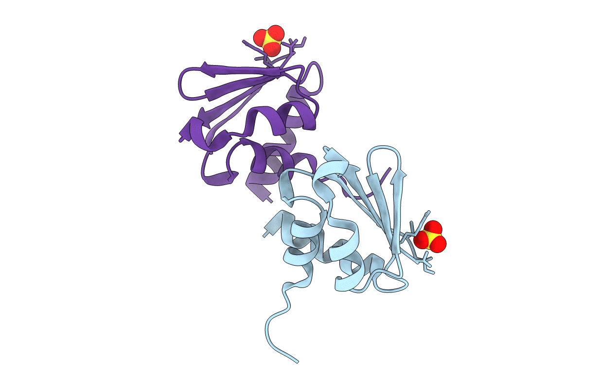
Crystal structure of EIIB
Biological Source:
Source Organism:
Host Organism:
PDB Version:
Deposition Date:
2007-12-18
Release Date:
2008-11-04
Method Details:
Experimental Method:
Resolution:
1.65 Å
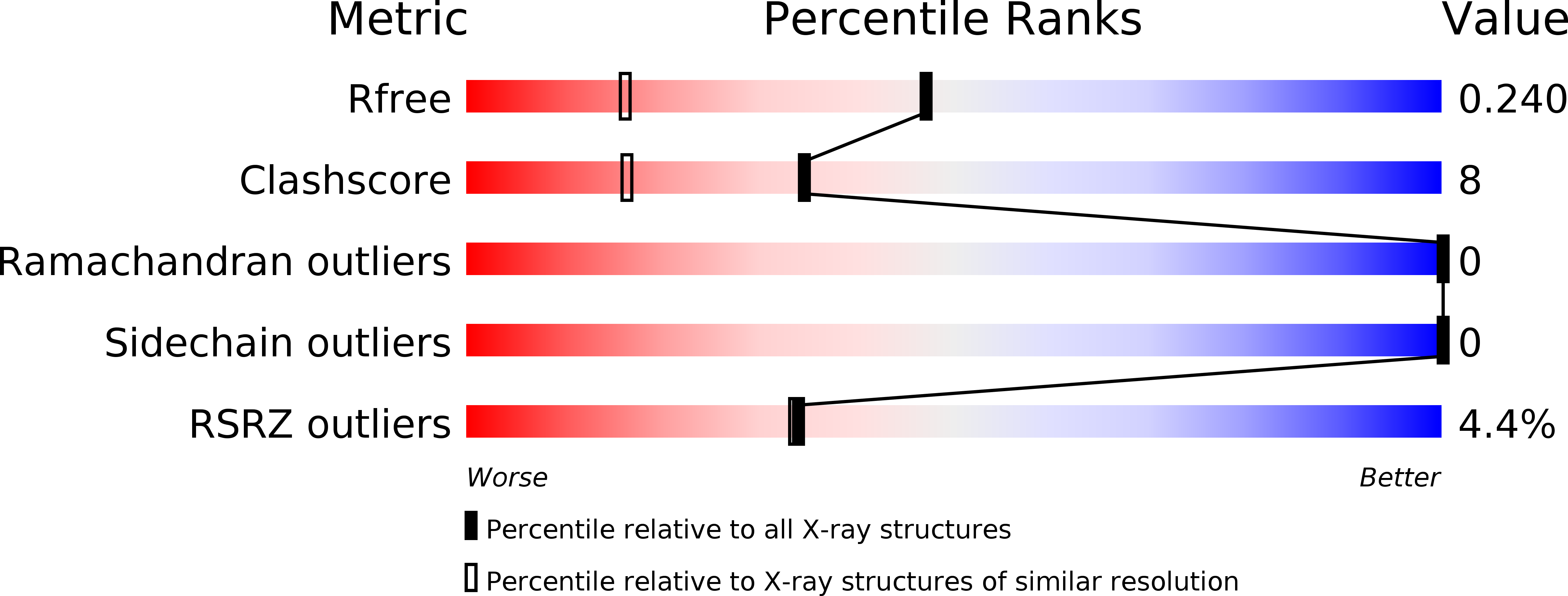
R-Value Free:
0.24
R-Value Work:
0.21
R-Value Observed:
0.21
Space Group:
C 2 2 21
Macromolecular Entities
Polymer Type:polypeptide(L)
Description:Glucose-specific phosphotransferase enzyme IIB component
Chain IDs:A, B
Chain Length:82
Number of Molecules:2
Biological Source:Escherichia coli
Modified Residue
Compound ID Chain ID Parent Comp ID Details 2D Image
MSE A MET SELENOMETHIONINE
Ligand Molecules
Primary Citation
Analyses of Mlc-IIBGlc interaction and a plausible molecular mechanism of Mlc inactivation by membrane sequestration.
Proc.Natl.Acad.Sci.Usa 105 3751 3756 (2008)
PMID: 18319344 DOI: 10.1073/pnas.0709295105

Abstact

In Escherichia coli, glucose-dependent transcriptional induction of genes encoding a variety of sugar-metabolizing enzymes and transport systems is mediated by the phosphorylation state-dependent interaction of membrane-bound enzyme IICB(Glc) (EIICB(Glc)) with the global repressor Mlc. Here we report the crystal structure of a tetrameric Mlc in a complex with four molecules of enzyme IIB(Glc) (EIIB), the cytoplasmic domain of EIICB(Glc). Each monomer of Mlc has one bound EIIB molecule, indicating the 1:1 stoichiometry. The detailed view of the interface, along with the high-resolution structure of EIIB containing a sulfate ion at the phosphorylation site, suggests that the phosphorylation-induced steric hindrance and disturbance of polar intermolecular interactions impede complex formation. Furthermore, we reveal that Mlc possesses a built-in flexibility for the structural adaptation to its target DNA and that interaction of Mlc with EIIB fused only to dimeric proteins resulted in the loss of its DNA binding ability, suggesting that flexibility of the Mlc structure is indispensable for its DNA binding.

Legend

Protein

Chemical

Disease

Primary Citation of related structures